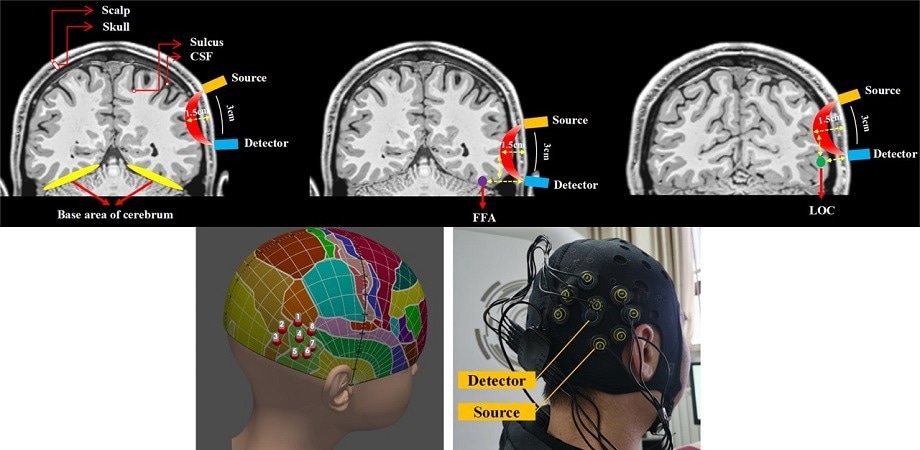
Not only is the brain the most intricate organ in the human body, but it is also one of the hardest to examine. Measuring neural activity in awake persons performing controlled tasks is essential to understanding the roles and interactions between various brain areas. Nevertheless, the most precise measurement tools are intrusive, which severely restricts their application on human subjects in a healthy environment.
 Functional near-infrared spectroscopy (fNIRS) can be performed using a portable emitter and detector placed on the scalp near a region of interest in the brain. By emitting and sensing light at specific frequencies, one can use fNIRS to detect changes in hemoglobin concentration, which correspond to brain activity. A transcranial brain atlas aids 3D localization for configuring optimal target channels. Image Credit: Chai, P. Zhang
Scientists have developed creative methods to detect brain activity in a safe and noninvasive manner to get over this significant impediment. One well-known example is functional magnetic resonance imaging (fMRI), which maps variations in cerebral blood flow using radio waves and strong magnetic fields.
The size and expense of the required equipment is a significant disadvantage of fMRI, preventing its wider use in clinical and laboratory settings. Thankfully, an alternative method known as functional near-infrared spectroscopy (fNIRS) has been gaining popularity.
This technique measures localized variations in hemoglobin content, which are correlated with brain activity, by applying a light source and detector to the scalp. Notwithstanding its benefits, which include portability and simplicity, the full potential of fNIRS is still untapped in many regions of the brain.
fNIRS has been utilized in earlier research to identify brain activity in the ventral visual pathway, but no assessment has been made of the technology's viability, ecological validity, or desirability of the signal found.
In light of this, a research team led by Professors Minghao Dong of Xidian University in China and Chaozhe Zhu of Beijing Normal University set out to investigate the efficacy of functional non-invasive retinal scanning (fNIRS) in assessing brain activity in the lateral occipital complex (LOC) and fusiform face area (FFA), two crucial areas in the ventral visual pathway. The Gold Open Access journal Neurophotonics published their findings.
Understanding the roles played by the LOC and FFA is helpful in understanding the experiments. The LOC's neurons are engaged in processing information regarding the forms and shapes of objects, which is important for object recognition.
The FFA, on the other hand, specializes in facial processing and recognition. The LOC is significantly closer to the scalp than the FFA. The scientists reasoned that this area was a better candidate for successful fNIRS measurements than the LOC.
The researchers gathered 63 adult participants to test this theory; 35 of them were included in the current study, and the remaining 28 were included in a follow-up study, the findings of which coincided with the current study but were not reported in the article. The group used a portable device to perform fNIRS measurements while carrying out a number of object and face recognition tasks.
The intention was to test if, during the experiment, photographs of faces or objects that the participant had previously seen would cause the appropriate brain region to become active. Notably, the researchers used a tool known as the "transcranial brain atlas," which they had created in a prior study, to figure out where the sensors on the instrument should be placed in relation to each particular patient.
Furthermore, the present study offers important new information by showing that LOC activity can be measured simply by aligning the target channel with the target coordinates; this eliminates the requirement for extra auxiliary channels surrounding the target coordinates.
“According to our findings, the LOC target channel selectively activates in reaction to objects, whereas the FFA target channel does not. The most likely explanation is that the depth at which the FFA is located exceeds the detection threshold of fNIRS, and the LOC region appears to be a suitable target for fNIRS-based detection, despite the fact that fNIRS detection has limitations in collecting FFA activity
Minghao Dong, Professor, Xidian University, China.
All things considered, the research team's efforts are a step in the right direction toward improved methods for studying the brain. Dong concludes, “Our results help understand the feasibility of fNIRS for practical applications. To the best of our knowledge, this work is the first to examine the viability of the technique in monitoring cortical activity within the ventral visual pathway.”
Additional developments in fNIRS technology may open up new possibilities for neuroenhancement devices and provide useful, affordable diagnostics for a number of brain illnesses. One could improve particular cognitive abilities or aid in the treatment of neurological disorders with the use of such devices.
Journal Reference:
Chai, Z., et al., (2024). Feasibility study of functional near-infrared spectroscopy in the ventral visual pathway for real-life applications, Neurophotonics. doi.org/10.1117/1.NPh.11.1.015002.