Oct 24 2012
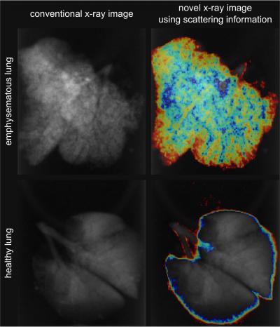
Chronic obstructive pulmonary disease (COPD) is considered the fourth most common cause of death in the United States. Usually the precursor to this life-threatening lung disease is a chronic bronchitis. Partially destroyed alveoli and an over-inflation of the lungs, known as emphysema, are serious side effects. However, the subtle differences in the tissue are barely discernable in standard X-ray images.
 A combination of dark-field and conventional transmission information allows for a clear distinction of healthy versus emphysematous tissue and an assessment of the regional distribution of the disease. From such images, a doctor might in future not only see if a patient is diseased but also which parts of the lung are affected and how much. Credit: Simone Schleede / Technische Universitaet Muenchen
In addition to the conventional X-ray images, the Munich scientists analyzed the radiation scattered by the tissue. From these data they calculated detailed images of the lungs of the investigated mice. Using such images, physicians can see not only if a patient is diseased but also how strongly which parts of the lung are affected.
"Especially in early stages of the disease, identification, precise quantification and localization of emphysema through the new technology would be very helpful", says Professor Maximilian Reiser, head of the Institute for Clinical Radiology at Ludwig-Maximilians-University Munich. "We hope that one day this technology will improve COPD diagnosis and therapy, while avoiding the higher radiation exposure associated with high-resolution CT".
The procedure has been developed as part of the research work of the Cluster of Excellence Munich-Centre for Advanced Photonics (MAP) by physicists from the Technische Universitaet Muenchen (TUM), physicians at the Ludwig-Maximilians-University Munich (LMU) and the Comprehensive Pneumology Center (CPC) of the Helmholtz Zentrum Muenchen.
For their experiments, the researchers used the Compact Light Source, a compact synchrotron radiation source of Lyncean Technologies Inc. (USA). In the future the Center for Advanced Laser Applications (CALA), a joint project of TUM and LMU on the Research Campus Garching, will develop new laser-driven x-ray sources.
In parallel, the research group led by Franz Pfeiffer, professor for Biomedical Physics at the Technische Universitaet Muenchen, works on the improvement of the x-ray scattering analysis to pave the way for its use with conventional X-ray machines.