Bo Huang, an assistant professor at Department of Pharmaceutical Chemistry and Department of Biochemistry and Biophysics at the University of California San Francisco, and Lei Zhu, an assistant professor at George W. Woodruff School of Mechanical Engineering of the Georgia Institute of Technology, have developed a new technique that enables a super-resolution microscope to perform live-cell imaging.
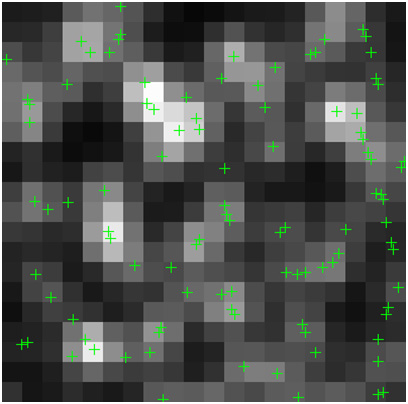
 The image shows single-molecule identification. The green cross signs show the locations of single molecules using the super resolution technique. (Image Courtesy of Lei Zhu and Bo Huang)
According to Zhu, the discovery of super-resolution fluorescence microscopy techniques such as photoactivated localization microscopy (PALM) and stochastic optical reconstruction microscopy (STORM) has overcome the diffraction limit of light microscopes. These techniques depend on the ability to record the emission of light from a sample’s single molecule. STORM/PALM detects the position of designated molecules utilizing probe molecules that are capable of switching between an invisible and a visible state. A structure can be clearly defined using these positions.
The new technique developed by Huang and Zhu builds on global optimization utilizing compressed sensing, which eliminates the estimation or assumption of the number of molecules in an image. The researchers demonstrated that compressed sensing is able to work with molecules of higher densities when compared to competitive technologies and illustrated fluorescent protein-labeled microtubules’ live cell imaging with a temporal resolution of three seconds.
Zhu informed that this technique can now be used to provide a large field view utilizing super-resolution microscopy with a temporal resolution of seconds or sub-seconds to observe several cellular processes in motion to better understand the life of a cell. The novel technique provides unprecedented temporal resolution and required spatial resolution to take images of dynamic single cellular structures. With this method, researchers can now find answers for new biological questions.
According to the researchers, this new technique also enables scientists to explore a vesicle’s active transports and other payloads within cells. The study findings have been reported in the Nature Methods journal.