Dec 23 2010
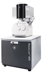
FEI announced that it has introduced a new scanning electron microscope (SEM) that is ideal for life sciences imaging applications. The Magellan SEM allows cell biologists and life sciences researchers to view a cell’s overall organization in its fully hydrated position.
The microscope incorporates a cryogenic sample preparation and provides an optimized process in order to deliver enhanced imaging and analysis results.
 Magellan SEM
The device generates subnanometer imaging in food sciences, nutrition, agricultural and plant applications at low accelerating voltages. The combination of high-resolution power, low-voltage imaging and cryogenic sample handling and preparation is crucial for imaging delicate biological systems such as plants, viruses and cells.
Cryogenic sample handling and preparation are important in life sciences applications in order to avoid adverse chemical drying procedures. The Magellan cryo workflow begins with a quick freezing procedure called vitrification. The process solidifies the water without creating any visible crystals.
When the vitrified sample is moved to the vacuum chamber, the Magellan SEM delivers imaging resolution in the entire range of accelerating voltages, beginning from 1 kV to 30 kV. The SEM functions at low voltages, accelerates the imaging signal’s surface specificity, enhances the resolution on light materials, and decreases the damage to fragile biological samples.