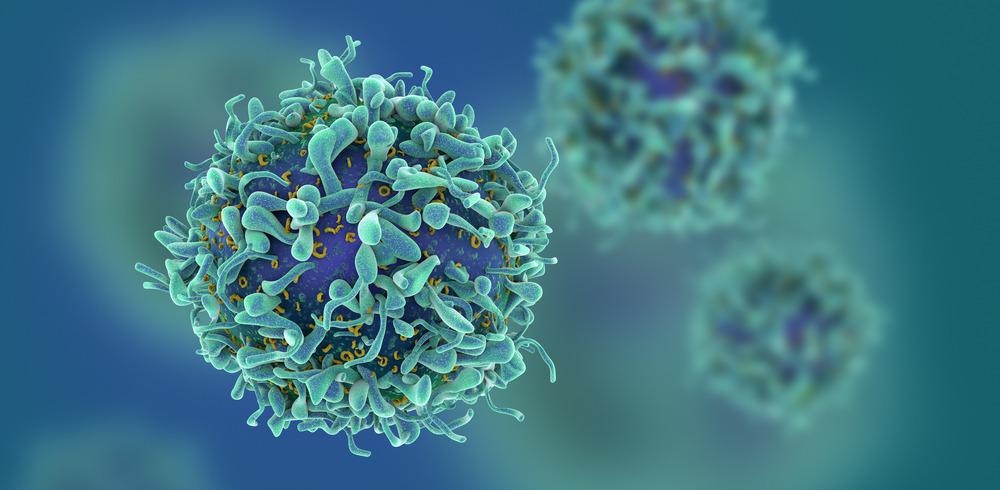
Photodynamic therapy (PDT) is a type of therapy that involves the use of light to activate a drug to trigger a cytotoxic effect. PDT has been used with great success in the treatment of a variety of cancer types, particularly those that are resistant to chemotherapy, as well as for the treatment of skin diseases such as acne and inflammatory rosacea.1

Image Credit: fusebulb/Shutterstock.com
One general model of how PDT works involves the formation of triplet states in the photosensitizer. The photosensitizer is the drug used that typically has a very strong light absorption in the ‘biological window’ – approximately 650 – 1350 nm. This is the region where skin and other tissues are reasonably transparent to the incident radiation.
Once the photosensitizer has been irradiated using a suitable laser pulse, it undergoes electronic relaxation. Most photosensitizers are designed so that, as part of this relaxation process, many molecules relax via electronic configurations that involve long-lived triplet states. Molecules in triplet states can then act as photosensitizers to produce radical oxygen species that can destroy any cancerous cells.2
There are now several generations of clinically approved photosensitizers for PDT.3 Many are used in routine medical treatments and there is a constant effort for the design and production of new drug species.2 However, it is a challenging task to find new molecules that show the required cytotoxicity.
Light Matters
While the biological window for light is typically considered to be 650-1350 nm, the transmission efficiency is significantly greater for longer infrared wavelengths. Because of this, photosensitizer development has focused on creating compounds with very efficient absorption in the infrared region. Although the doses of light used in current PDT treatments are not harmful, there is a limit to how much light radiation can be used before skin damage and irritation becomes a serious risk.1
An alternative approach has been to use multi-photon absorption processes to excite the photosensitizer rather than a single photon. Most molecules have lower two-photon absorption cross-sections than for one-photon, but for those with sufficiently efficient two-photon absorption, the same amount of energy can be delivered as a single higher-energy photon by using multiple long-wavelength photons that have higher transmission.4 This helps overcome some of the issues with tissue transmissions but as this is a non-linear optical process, it often requires laser sources with high peak powers.
Tackling treatment of deep-seated tumors is still a challenge for PDT. Skin cancers and diseases can be treated very effectively as there is little concern about the transmission issue. However, deep-seated tumors may require the delivery of light via fiber optics, which also implies some amount of surgical treatment.
X-Ray Transmission
Recently, a new generation of photosensitizers has been developed that can be excited with X-ray radiation.5 Except for the densest tissues in our bodies, such as bones, our bodies are mostly transparent to high-energy X-ray radiation. In clinical mimics, these copper-based photosensitizers showed efficient singlet oxygen generation for tumor destruction, but also some ability to inhibit the migration and proliferation of cancerous cells.5 No obvious toxicities were found in cell studies, making these copper complexes a highly appealing candidate for further study.
A common method for the evaluation of potential complexes for PDT is to compare the ‘dark’ and ‘light’ toxicities. This is because an ideal PDT candidate will show no toxicity under dark conditions as the photosensitizer has not been activated. Low dark toxicity reduces the risk of unwanted side effects, particularly while the drug is transported to the tumor location. Ideally, the drug will show good take-up only at targeted tumor sites as the generation of radical oxygen species can be damaging to healthy and cancerous cells alike.
The copper complexes showed low dark cytotoxicity, except at very high photosensitizer concentrations. This is a common issue with metal-based photosensitizers as, although the presence of metal often enhances the yields of radical oxygen production, many of these elements are cytotoxic themselves at high concentration.
When irradiated with X-rays from a clinical X-ray source, the cytotoxicity was significantly enhanced. This also led to some amount of promotion of anti-tumor immune responses. A challenging aspect of cancer treatment is balancing damage to healthy tissue with complete eradication of cancerous cells. By promoting immune responses, this type of PDT treatment may prove even more effective overall as any cancerous cells not destroyed in the initial cell death triggered by the formation of reactive oxygen species can later be destroyed by the immune response. This is also thought to be how these copper complexes can inhibit tumor formation.
The development and trial of PDT compounds that are photoactive by X-rays rather than visible or infrared light is an exciting development for increasing the treatment possibilities of the technique.
References and Further Reading
- Niculescu, A. (2021). Photodynamic Therapy — An Up-to-Date Review. Applied Sciences, 11, 3623. https://doi.org/10.3390/app11083626
- Monro, S., Colón, K. L., Yin, H., Roque, J., Konda, P., Gujar, S., Thummel, R. P., Lilge, L., Cameron, C. G., & McFarland, S. A. (2019). Transition Metal Complexes and Photodynamic Therapy from a Tumor-Centered Approach: Challenges, Opportunities, and Highlights from the Development of TLD1433. Chemical Reviews, 119(2), 797–828. https://doi.org/10.1021/acs.chemrev.8b00211
- Detty, M. R., Gibson, S. L., & Wagner, S. J. (2004). Current clinical and preclinical photosensitizers for use in photodynamic therapy. Journal of Medicinal Chemistry, 47(16), 3897–3915. https://doi.org/10.1021/jm040074b
- McKenzie, L. K., Bryant, H. E., & Weinstein, J. A. (2019). Transition metal complexes as photosensitisers in one- and two-photon photodynamic therapy. Coordination Chemistry Reviews, 379, 2–29. https://doi.org/10.1016/j.ccr.2018.03.020
- Chen, X., Liu, J., Li, Y., Pandey, N. K., Chen, T., Wang, L., Amador, E. H., Chen, W., Liu, F., Xiao, E., & Chen, W. (2021). Study of copper-cysteamine based X-ray induced photodynamic therapy and its effects on cancer cell proliferation and migration in a clinical mimic setting. Bioactive Materials, 7(April 2021), 504–514. https://doi.org/10.1016/j.bioactmat.2021.05.016
Disclaimer: The views expressed here are those of the author expressed in their private capacity and do not necessarily represent the views of AZoM.com Limited T/A AZoNetwork the owner and operator of this website. This disclaimer forms part of the Terms and conditions of use of this website.