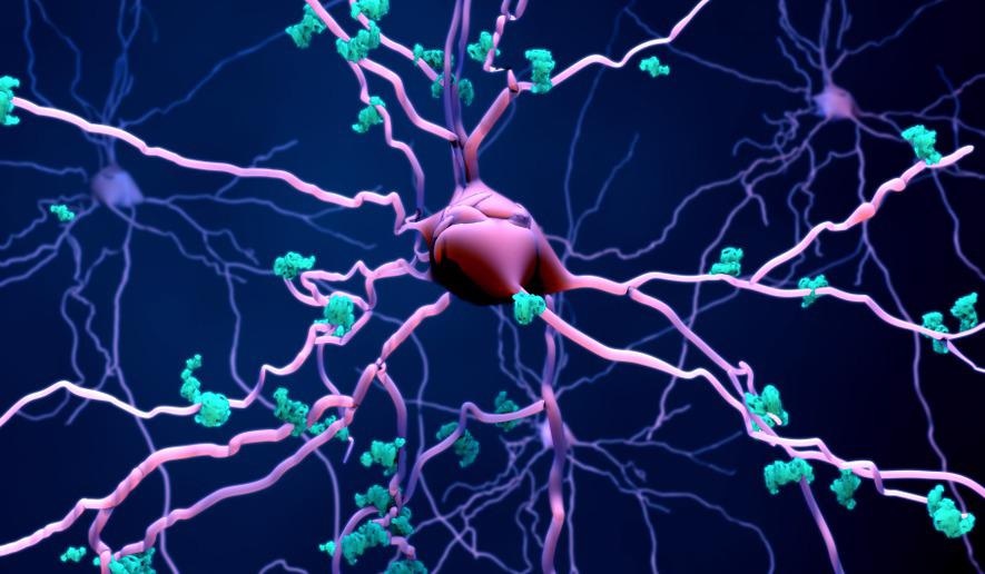
Electron microscopy coupled with fluorescence has validated the efficacy of a new technique designed to genetically tag proteins in a predetermined neural location. The method, created by researchers at Northwestern University and the University of Pittsburgh, will help scientists gain a deeper understanding of neurological illnesses and potentially develop new therapeutic approaches based on this knowledge.

Image Credit: Design_Cells/Shutterstock.com
The Link Between Proteins and Neurological Illness
Proteins are the second most prevalent type of matter in the brain, second to water. Therefore, it is unsurprising that they hold the key to understanding many facets of brain function and brain disease.
Research has shown that several neurological illnesses, including Parkinson's disease (PD), Alzheimer’s disease (AD), multiple system atrophy (MSA), and dementia with Lewy bodies (DLB) are associated with abnormal protein production and build up. For example, accumulations of the α-synuclein protein have been found in the brains of people with PD, MSA, and DLB. Additionally, the amyloid precursor protein (APP), β- amyloid (Aβ), and tau protein have been implicated in the initiation and progression of AD.
It is vital to investigate the relationship between protein activity and these diseases to understand their underlying cause and pathology. In doing so, future potential therapeutic options can be explored. With this method, scientists will be able to gain vital insights into how neurons in the brain function. This knowledge will be integral to furthering our understanding of neurological diseases and hopefully enable the development of new therapeutic approaches.
Tagging Proteins in the Living Brain
In a study published in August 2021 in the journal Nature Communications, a team of US scientists describes how they designed a virus capable of directing an enzyme to a specific location within the brain of a living mouse. The enzyme, derived from soybeans, demonstrated its ability to generally tag proteins in its surrounding area once it reaches its predetermined location.
Electron microscopy was used alongside a fluorescence technique to validate the novel method. APEX2, an engineered peroxidase, was virally induced to tag proteins in the neurons located within the striatum. Transmission electron microscopy distinguished the nuclear exporting sequence (NES) and membrane (LCK) constructs via the examination of localization patterns in brain slices. The results revealed a snapshot of the entire proteome inside the living neurons of the mice.
While similar work has been conducted previously, it had been limited to studying cells in cultures. The results found in cellular cultures are often different from those displayed in living cells, as the two function differently in these distinct environments.
Overcoming this, researchers have established a method that effectively tags proteins and their neighbors, revealing how proteins within a target location work and interact with each other in living cells.
Here, scientists used the virus to deliver the 'message' of the enzyme, acting as a vessel for information. The scientists also sent a fluorescent protein to reveal which neurons had been tagged in a separate message. Illuminated neurons represented those that expressed the enzyme.
The field of genetic targeting has rapidly developed over the years, allowing biology and neuroscience disciplines, among other scientific sectors, to transform dramatically.
Developments in protein targeting, however, have lagged. Although gene and RNA sequences can be amplified to identify the amino acids they code for, the method cannot be applied to proteins. Instead, proteins must be split into and then placed back together, which is time-consuming and prone to errors.
Now, with the novel approach established by researchers at Northwestern University and the University of Pittsburgh, the proteome of a range of neural circuits can be mapped with high levels of precision and specificity. Proteins in specific circuitries will even be able to be quantified so scientists can establish the number of different types of proteins in various areas of the brain.
Furthering our Knowledge of Neurological Illnesses
With the new approach validated, researchers can apply it to various mouse models of neurological illness to develop a deeper understanding of such diseases, including AD, PD, MSA, and DLB.
The scientists involved in developing the novel method are interested in extended the approach. Aspirations center around determining the biochemical modifications of neuronal proteins that are associated with specific types of brain activity, such as under the influence of neuroactive drugs.
The new method will likely help further our knowledge of neurological diseases and establish new therapeutic options. Given the limited treatment options currently available for such conditions, this new technique may help to improve the quality of life of thousands of people living with neurological diseases.
References and Further Reading
Dumrongprechachan, V., Salisbury, R., Soto, G., Kumar, M., MacDonald, M. and Kozorovitskiy, Y., (2021) Cell-type and subcellular compartment-specific APEX2 proximity labeling reveals activity-dependent nuclear proteome dynamics in the striatum. Nature Communications, 12(1). Available at: https://doi.org/10.1038/s41467-021-25144-y [Acccessed August 2021]
Lam, S., Martell, J., Kamer, K., Deerinck, T., Ellisman, M., Mootha, V. and Ting, A., (2014) Directed evolution of APEX2 for electron microscopy and proximity labeling. Nature Methods, 12(1), pp.51-54. Available at: https://doi.org/10.1038/nmeth.3179 [Acccessed August 2021]
Penke, B., Bogár, F., Paragi, G., Gera, J. and Fülöp, L., (2019) Key Peptides and Proteins in Alzheimer’s Disease. Current Protein & Peptide Science, 20(6), pp.577-599. Available at: https://doi.org/10.2174/1389203720666190103123434 [Acccessed August 2021]
Snyder, H. and Wolozin, B., (2004) Pathological Proteins in Parkinson's Disease: Focus on the Proteasome. Journal of Molecular Neuroscience, 24(3), pp.425-442. Available at: https://doi.org/10.1385/jmn:24:3:425 [Acccessed August 2021]
Northwestern University. (2021) New technique identifies proteins in the living brain: Tool could help researchers better understand brain diseases and potential treatments. [online] ScienceDaily. Available at: www.sciencedaily.com/releases/2021/08/210811091541.htm [Acccessed August 2021]
Disclaimer: The views expressed here are those of the author expressed in their private capacity and do not necessarily represent the views of AZoM.com Limited T/A AZoNetwork the owner and operator of this website. This disclaimer forms part of the Terms and conditions of use of this website.