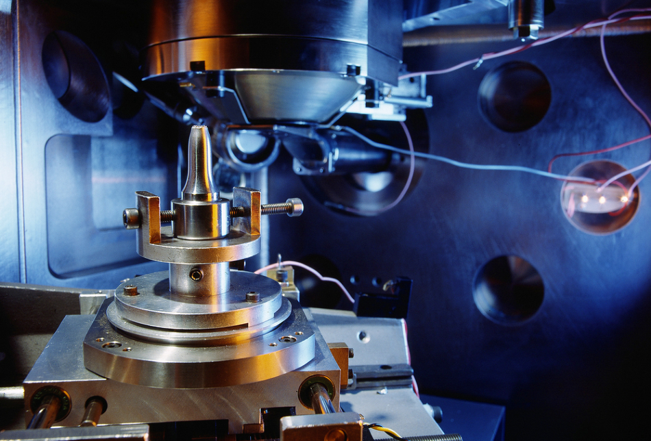
Image Credit: Bildagentur Zoonar GmbH/Shutterstock.com
Quantum physics began to revolutionize the way we understand the universe in its smallest possible spatial dimensions from the 1910s. Electron microscopy developed in the early twentieth century as one of its foremost experimental applications. This article explains how electron microscopy works.
Electrons and Photons: A Difference of Scale
Electron microscopes manipulate electrons, which are quantum particles, to illuminate and magnify the specimen being observed. They are capable of 10,000,000 times magnification at resolutions as precise as only 50 picometers (one trillionth of a meter).
This level of magnification, which is much greater than traditional optical microscopes, is achieved due to the shorter wavelength of electrons compared to photons. Because the wavelength of a photon is between 400 and 700 nm, objects or details smaller than this can never be accurately imaged in a microscope that uses light to illuminate the specimen. Electrons, on the other hand, have a wavelength of approximately 4 pm.This means that magnification using electrons can be much greater without losing resolution.
The difference between photons and electrons could be illustrated as the difference between a fine tip pen and a large marker: the large marker will never be able to achieve the same level of detail in a drawing as a fine tip pen.
Using Electromagnetism to Create a Lens
Traditional optic microscopy bends a source of emitted light through a refractive glass lens. This bending has the effect of magnifying the specimen, as light returning from the specimen to the observer is effectively spread out evenly by the curve of the lens.
Electron microscopy replaces the light source with a source of emitted electrons. These quantum particles – which carry the electronic charge in an atom, or carry electricity through conductive materials – are sent through a series of coil-shaped electromagnets towards the specimen. This has the effect of changing the quantum state of electrons by creating an electron-optical lens – an electromagnetic field shaped like the glass lens found in an optical microscope.
The image of the specimen object or detail behind this electron lens can then be recorded in a photograph or in a computer, and will be magnified to the high levels discussed above.
Different Types of Electron Microscope
There are different applications of the lensing effect of electromagnetic coils for electron microscopy. In each, the quantum states of electrons change in a different way.
The transmission electron microscope (TEM) was the first electron microscope developed. In a TEM, electrons are fired in a powerful beam of extremely high frequency through the specimen material. Here the state of electrons is of a high-frequency wave, concentrated and focused by electromagnetic coils.
After passing through the specimen, a third electromagnetic coil magnifies the electrons like a lens and this magnified image is detected by a fluorescent screen.
In a scanning electron microscope (SEM), electron particles are fired into a series of electromagnetic coils which create an intense beam, similar to that in the TEM. This beam of electrons bounces off of the specimen surface, and is scanned across the specimen by further coils.
These returning electrons are registered by a detector, and a magnified image can be produced.
The Changing Quantum States of Electrons in an Electron Microscope
These modes of electron microscopy work due to the dual quantum states of electrons which, like photons, can be both waves and particles. The discovery of this duality in light was the basis of modern quantum physics, and the ability of electrons’ quantum states to change from wave to particle, or be both at once, continues to fascinate experimental and theoretical physicists today.
References and Further Reading
Erni, R., Rossell, M.D., Kisielowski, C. and Dahmen, U. (2009). Atomic-Resolution Imaging with a Sub-50-pm Electron Probe. Physical Review Letters, 102(9). DOI:10.1103/PhysRevLett.102.096101
Freundlich, M.M. (1963). Origin of the Electron Microscope. Science, 142(3589), pp.185–188.Freundlich, M.M. (1963). Origin of the Electron Microscope. Science, 142(3589), pp.185–188.
Disclaimer: The views expressed here are those of the author expressed in their private capacity and do not necessarily represent the views of AZoM.com Limited T/A AZoNetwork the owner and operator of this website. This disclaimer forms part of the Terms and conditions of use of this website.