Jan 23 2010
A team of researchers from the Max Planck Institute of Biochemistry, in Germany, led by the Spanish physicist Rubén Fernández-Busnadiego, has managed to obtain 3D images of the vesicles and filaments involved in communication between neurons. The method is based on a novel technique in electron microscopy, which cools cells so quickly that their biological structures can be frozen while fully active.
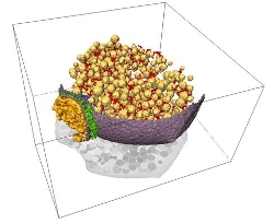
 Visualización tridimensional de la sinapsis mediante tomografía electrónica: vesículas sinápticas (amarillo), membrana celular (violeta), conectores entre vesículas (rojo), filamentos que anclan las vesículas a la membrana celular (azul marino), microtúbulo (verde oscuro), material del espacio sináptico (verde claro) y densidad postsináptica (naranja).
“We used electron cryotomography, a new technique in microscopy based on ultra-fast freezing of cells, in order to study and obtain three-dimensional images of synapsis, the cellular structure in which the communication between neurons takes place in the brains of mammals” Rubén Fernández-Busnadiego, lead author of the study which features on the front cover of this month’s Journal of Cell Biology and a physicist at the Max Planck Institute of Biochemistry, in Germany, tells SINC.
During synapsis, a presynaptic cell (emitter) releases neurotransmitters to another post-synaptic one (recipient), generating an electric impulse in it, thereby allowing nervous information to be transmitted. During this study, the researchers focused on the tiny vesicles (measuring around 40 nanometres in diameter), which transport and release the neurotransmitters from the presynaptic terminals.
“Thanks to the use of certain pharmacological treatments and the advanced 3D imaging analysis method we have developed, it is possible to observe the huge range of filamentous structures that are within the presynaptic terminal and interact directly with the synaptic vesicles, as well as to learn about their crucial role in responding to the electrical activity of the brain,” explains Fernández-Busnadiego.
The filaments connect the vesicles and also connect them with the active area, the part of the cellular membrane from which the neurotransmitters are released. According to the Spanish physicist, these filamentous structures act as barriers that block the free movement of the vesicles, keeping them in their place until the electric impulse arrives, as well as determining the ease with which they will fuse with the membrane.