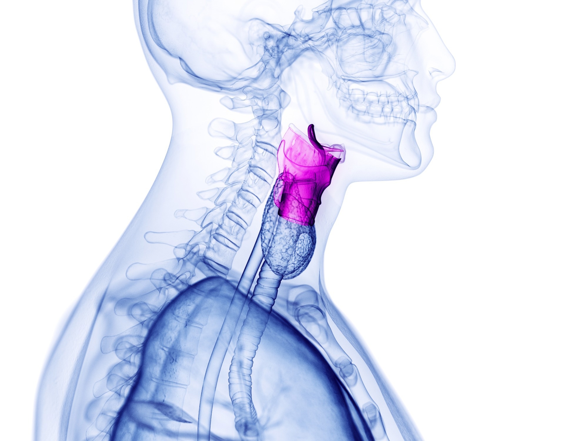
The larynx, sometimes referred to as the “voice box,” contains tiny, flexible flaps of tissue called the vocal folds, also known as vocal cords. These fragile, complex structures support breathing and swallowing, among other bodily processes. But most crucially, the vocal folds provide the ability to speak.

Image Credit: SciePro/Shutterstock.com
By manipulating the tension and form of these folds, humans can create a wide range of sounds. The mechanical characteristics of the vocal folds can, sadly, be impacted by a wide variety of disorders and abnormalities. These folds’ significant flexibility and viscosity fluctuations might cause voice loss or seriously impair the ability to speak.
Currently, laryngoscopy and videostroboscopy are used to diagnose all vocal fold disorders. These methods make it possible to see the vocal folds in operation, giving hints about any underlying conditions. However, a biopsy, a dangerous technique that could have unanticipated negative consequences on the voice, is now the only option to objectively assess the mechanical qualities of the vocal folds and arrive at a conclusive diagnosis.
A study team from Texas A&M University and the University of Southern California is working on a non-invasive method to examine the mechanical characteristics of the vocal folds to solve this problem.
The researchers presented Brillouin microspectroscopy, an emerging optical imaging technique with tremendous potential for the identification of vocal fold diseases, and its untapped potential in a new study published in the Journal of Biomedical Optics.
The suggested approach is founded on the idea of Brillouin scattering, an optical phenomenon where light interacts with sound waves in a material to produce mechanical vibrations. Depending on the elasticity of the material, this results in a slight shift in the frequency of light reflected from the material’s surface.
The researchers created a unique microspectroscopy system that was explicitly designed to study Brillouin scattering in vocal fold tissues. They measured samples of porcine vocal folds to validate their theory, and then only based on the Brillouin frequency shifts; they computed the elastic characteristics of the samples. After that, they made a comparison between the estimated values and those discovered in earlier research using traditional elasticity measurements.
Overall, the researchers discovered good agreement between the outcomes of the two assessment techniques. The team was able to precisely assess the stiffness variations between the superior and inferior vocal folds using the Brillouin microspectroscopy technique.
The observed Brillouin frequency shifts alone allowed them to distinguish different laryngeal substructures from the surrounding tissue: the superior vocal fold had the lowest shift, while the supraglottal wall tissue showed the highest shift.
The results of this study suggest that Brillouin microspectroscopy could someday be a useful tool for laryngologists. The suggested imaging platform could be made compatible with the advanced endoscopic machinery that professionals often use to examine vocal folds. This could pave the way for non-invasive, remote vocal fold pliability assessments and, consequently, new diagnostic criteria.
Journal Reference:
Cheburkanov, V., et al. (2023) Porcine vocal fold elasticity evaluation using Brillouin spectroscopy. Journal of Biomedical Optics. doi:10.1117/1.JBO.28.8.087002