 By Owais AliReviewed by Lexie CornerSep 25 2024
Biomedical optics has become a powerful and versatile field in modern medicine, providing non-invasive imaging, precise diagnostics, and targeted therapies that offer unparalleled insights into the human body. This article reviews the fundamental principles of biomedical optics and its key imaging and therapeutic applications.
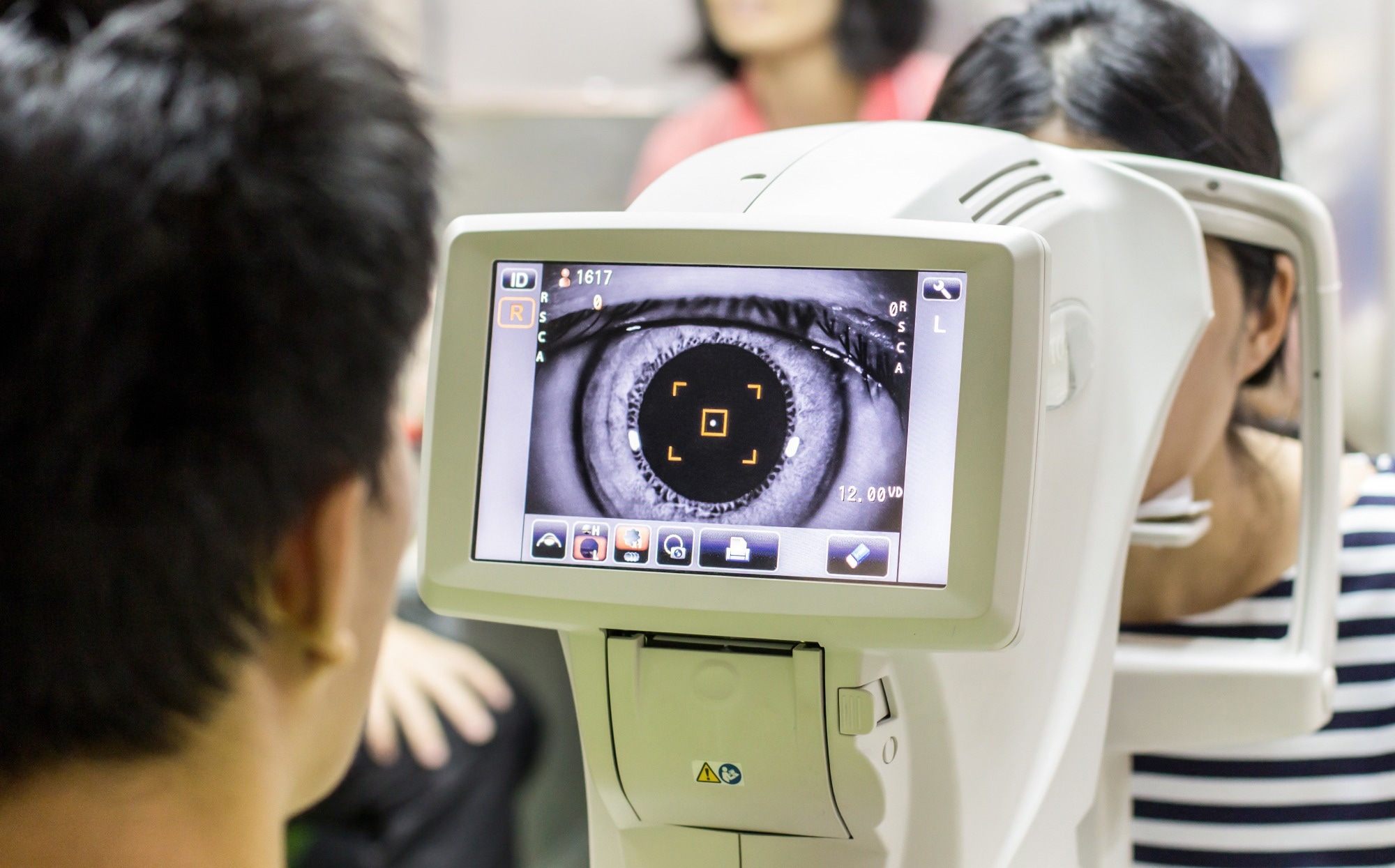
Image Credit: J.Thasit/Shutterstock.com
What is Biomedical Optics?
Biomedical optics is a multidisciplinary field focused on the interaction of light with biological systems, from organs to cells, to develop technologies for research and clinical applications. It uses optical principles to achieve high-resolution, real-time imaging and analysis of biological structures and processes.
This field has transformed medical diagnostics by introducing non-invasive and minimally invasive tools that improve early disease detection, treatment planning, and monitoring of therapeutic outcomes. Additionally, biomedical optics has facilitated the development of targeted treatments that reduce patient discomfort and recovery times compared to conventional methods.1
Key Principles of Light-Tissue Interaction
A key challenge in biomedical optics is understanding how light interacts with complex, inhomogeneous tissues. These interactions involve reflection, refraction, absorption, and multiple photon scattering, complicating accurate imaging and analysis.
Absorption occurs when photon energy is absorbed by tissue components, such as chromophores, resulting in a reduction in light intensity as it penetrates deeper into the tissue. The extent of absorption varies with wavelength, affecting how different wavelengths penetrate tissues, influencing tissue penetration depth and enabling the differentiation of tissue types based on their absorption spectra.
Scattering occurs when photons deviate from their original path due to interactions with tissue structures, such as cell membranes and the extracellular matrix. It can be categorized into single scattering, which happens in transparent tissues, or multiple scattering, which is more common in opaque tissues.
In opaque tissues, such as skin and brain, light experiences significant multiple scattering, causing the light beam to broaden and decay. This impacts the quality of imaging and the efficacy of light-based diagnostic techniques, making the understanding and modeling of scattering essential for improving optical imaging methods.
Refraction occurs when light passes through tissues with varying refractive indices, causing a change in its direction. This bending of light can affect imaging and diagnostic techniques by altering the perceived location and properties of tissue structures.2,3
Imaging Applications
One of biomedical optics’ most significant contributions to healthcare is its range of noninvasive imaging techniques. These methods enable early disease detection and precise monitoring, providing detailed anatomical insights that greatly enhance patient care.
OCT is a high-resolution imaging technique that uses light to capture real-time, cross-sectional images of biological tissues by measuring the time delay of backscattered light through interferometry. It provides an imaging depth of 1-2 mm in scattering tissues and a resolution between 1 and 10 µm.
OCT is primarily used in ophthalmology for non-invasive retinal imaging, but it also has applications in cardiology, dermatology, and other fields.4
Photoacoustic Tomography (PAT)
PAT combines optical absorption with ultrasonic wave generation to produce detailed images of tissues. A short-pulsed laser induces localized heating in tissue through thermoelastic expansion, generating ultrasonic waves that are captured and analyzed to create images based on the tissue's absorption properties.
This imaging technique is useful for visualizing blood vessels, assessing tissue oxygenation, and evaluating various tissue characteristics, making it valuable in fields such as vascular imaging and cancer detection.
Hyperspectral Imaging (HSI)
HSI is another non-invasive technique that captures spectral information across a range of wavelengths for each pixel in a 2D detector array, creating a three-dimensional hypercube. This dataset provides detailed spatial and spectral information, allowing for the detection of tissue abnormalities by analyzing changes in tissue absorption, fluorescence, and scattering.
It is commonly used for cancer detection, assessing diabetic ulcers, and monitoring cardiovascular conditions.2
Fluorescence Imaging
Fluorescence imaging uses fluorescent dyes to enhance contrast in biological tissues and cells by labeling specific structures with fluorophores that emit light upon excitation. This technique enables detailed visualization of cellular components and includes various methods such as fluorimetry, widefield microscopy, and fluorescence-lifetime imaging, offering high specificity and resolution for studying cellular processes, diagnosing diseases, and guiding surgeries.5,6
Therapeutic Applications
While imaging highlights the diagnostic capabilities of biomedical optics, its therapeutic applications have equally transformative potential.
Photodynamic Therapy (PDT)
PDT involves administering a photosensitizer (PS), a non-toxic drug or dye that accumulates in malignant or targeted tissues. After the incubation period, the area is exposed to red visible light (620–690 nm), which, in the presence of oxygen, generates reactive oxygen species (ROS) that induce cytotoxic effects and tissue destruction.
PDT is highly valued for its specificity; the PS accumulates in malignant tissues, ensuring that light activation primarily affects the diseased area.7
Laser Therapies
Laser therapy includes low-level laser therapy (LLLT) and high-intensity laser therapy (HILT), which employ photo-biomodulation to provide therapeutic effects. LLLT targets superficial tissues with energy outputs up to 500 mW, whereas HILT, with outputs greater than 500 mW, can penetrate deeper tissues and induce photothermal effects.
HILT has been shown to alleviate pain, enhance functional activity, and is particularly effective for musculoskeletal disorders like neck pain. In addition, it demonstrates superior efficacy in pain management and functional improvement compared to other non-invasive methods like ultrasound and TENS.8
Photothermal Therapy (PTT)
PTT uses light to generate localized heat in targeted tissues, typically using light-absorbing nanoparticles. Pathological cells are labeled with these nanoparticles, which increase heat generation upon exposure to near-infrared (NIR) light. This targeted heating enables precise destruction of tumor cells while minimizing damage to surrounding healthy tissues.
PTT has shown promise in treating various cancers by targeting tumors with minimal damage to adjacent healthy cells, and ongoing research continues to explore its efficacy and safety in clinical applications.9
Recent Trends and Advancements
AI-Enhanced Retinal Imaging
One of the most promising trends in biomedical imaging is the integration of artificial intelligence (AI), which enhances diagnostic precision by analyzing complex data, refining image processing, and accelerating workflows.
Recently, a study published in Communications Medicine by the National Institutes of Health researchers reported significant advancements in retinal imaging through the integration of artificial intelligence (AI) with adaptive optics (AO) and optical coherence tomography (OCT). This combination achieved a 100-fold increase in imaging speed and a 3.5-fold improvement in image contrast.
The researchers developed the parallel discriminator generative adversarial network (P-GAN), a deep learning algorithm trained on nearly 6,000 AO-OCT images, which efficiently de-speckles images and reduces processing time by approximately 100-fold compared to traditional methods.
This AI-driven approach enhances image quality and accessibility, making AO-OCT imaging more viable for routine clinical use and detailed studies of retinal diseases affecting the retinal pigment epithelium (RPE).10
A Cost-Effective, Non-Invasive Drug Detection Wearable Sensor
Another notable trend is the rise of wearable biosensors, which continuously monitor physiological parameters like heart rate and glucose levels, providing real-time health data for proactive management and early detection of potential issues.
A study published in ACS Applied Materials & Interfaces has proposed a highly sensitive wearable sensor that detects illegal drugs in sweat. This sensor offers a rapid, noninvasive, and cost-effective alternative to traditional drug testing methods.
Unlike traditional drug testing methods that involve complex extraction and chromatography, this sensor is applied as a sweat patch and requires no additional preparation. It uses surface-enhanced Raman scattering (SERS) to amplify the optical signal of narcotics, enabling rapid, real-time, non-invasive detection in just one minute.
With a production cost under 50 cents per unit, this sensor offers a cost-effective solution for anti-doping tests and other applications, addressing privacy concerns and providing a practical alternative for large-scale drug monitoring.11
Conclusion
Biomedical optics represents a dynamic and rapidly evolving field that has transformed our approach to diagnosing and treating medical conditions. As optical technologies advance, biomedical optics is set to play a central role in the future of medicine, offering more precise, personalized, and effective healthcare.
More from AZoOptics: Optical Filters: Exploring the Differences Between Absorption and Interference Filters
References and Further Reading
- Linköping University. (2024). Biomedical Optics. [Online] Linköping University. Available at: https://liu.se/en/research/biomedical-optics
- Keiser, G. (2016). Biophotonics. Springer. https://doi.org/10.1007/978-981-19-3482-7
- Tuchin, VV. (2003). Light-tissue interactions. Biomedical Photonics Handbook. 3-1. https://www.taylorfrancis.com/chapters/edit/10.1201/b17290-9/light%E2%80%93tissue-interactions-valery-tuchin
- Lahoti, HS., Jogdand, SD. (2022). Bioimaging: evolution, significance, and deficit. Cureus. https://doi.org/10.7759/cureus.28923
- Novanta Photonics. (2022). An Introduction to Fluorescence Imaging. [Online] Novanta Photonics. Available at: https://novantaphotonics.com/introduction-fluorescence-imaging/
- Sanderson, MJ., Smith, I., Parker, I., Bootman, MD. (2014). Fluorescence microscopy. Cold Spring Harbor protocols. https://doi.org/10.1101/pdb.top071795
- Robertson, CA., Evans, DH., Abrahamse, H. (2009). Photodynamic therapy (PDT): a short review on cellular mechanisms and cancer research applications for PDT. Journal of Photochemistry and Photobiology B: Biology. https://doi.org/10.1016/j.jphotobiol.2009.04.001
- Xie, YH., Liao, M. X., Lam, FM., Gu, YM., Fernando, WHA., Liao, LR., Pang, M. Y. (2023). The effectiveness of high-intensity laser therapy in individuals with neck pain: a systematic review and meta-analysis. Physiotherapy. https://doi.org/10.1016/j.physio.2023.07.003
- Gellci, K., Mehrmohammadi, M. (2014). Photothermal Therapy. Encyclopedia of Cancer. https://doi.org/10.1007/978-3-642-27841-9_7097-1
- Das, V., et al. (2024). Revealing speckle obscured living human retinal cells with artificial intelligence assisted adaptive optics optical coherence tomography. Communications Medicine. https://doi.org/10.1038/s43856-024-00483-1
- Koh, EH., et al. S. (2021). A wearable surface-enhanced Raman scattering sensor for label-free molecular detection. ACS Applied Materials & Interfaces. https://doi.org/10.1021/acsami.0c18892
Disclaimer: The views expressed here are those of the author expressed in their private capacity and do not necessarily represent the views of AZoM.com Limited T/A AZoNetwork the owner and operator of this website. This disclaimer forms part of the Terms and conditions of use of this website.