Professor Milena D’Angelo’s research group at the Dipartimento Interateneo di Fisica, Università degli Studi di Bari, has devised an innovative light-field microscopy technique for volumetric imaging. Published in Nature - Scientific Reports, its experiments show how a novel technique termed correlation light-field microscopy (CLM), which uses the correlation between two light beams to obtain volumetric imaging, is only constrained in resolution by light diffraction.

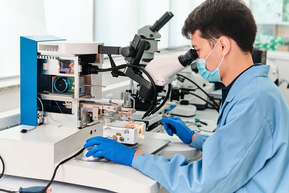
Study: Light-field microscopy with correlated beams for high-resolution volumetric imaging. Image Credit: Andrey Yershov/Shutterstock.com
Microscopy has long struggled to quickly image three-dimensional materials at the diffraction limit. Numerous attempts have been made to solve the necessity for quick imaging of vast volumes with fast acquisition speeds to study dynamic biological processes. All of these methods have resulted in various tradeoffs.
Technological improvements to acquire better images in this area include:
- Multi-focus multiplexing
- Fast two-photon microscopy
- Fast stimulated emission depletion microscopy (STED)
- Light-sheet illumination
- Depth-focused scanning with tunable lenses
Light-field microscopy (LFM) is a technique that has emerged with the most promise.
What is Light-Field Microscopy (LFM)?
The term "light field" is used in computer vision and photography to refer to the distribution of radiance from a source or the amount of light the object emits as a function of both position and view direction.
In a traditional widefield microscope, directional (angular) information is lost because all rays originating from a point in the object within the objective lens' acceptance cone (defined by its numerical aperture) are concentrated to a single point on the camera sensor.
LFM approaches aim to simultaneously acquire 4D radiance data, providing volumetric information about the object.
Light-field imaging has made it possible to concentrate out-of-focus areas of three-dimensional samples during post-processing by identifying light's spatial distribution and propagation direction in a single exposure. Stacking refocused planes at various distances increases the depth of field (DOF) within the imaged volume.
When using a lens for observation, an object is best seen when it is in the focus point of the lens. There is a range of tolerance in which the object can still be seen even if the distance between the lens and the subject is slightly offset. The depth-of-field (DOF) is the term for this range.
In its traditional application, light-field imaging is limited by the compromise between the resolution and DOF. This barrier is particularly unfavorable in the microscopic pursuit since the sought-after high resolution severely restricts the DOF. Repetitive scanning and accumulation of large data are required to characterize a thick sample.
In this paper, Prof. D’Angelo’s team describes developing and testing a novel technique for performing light-field microscopy with diffraction-limited resolution through experimental demonstration.
Correlation light-field microscopy is a novel method that can surpass conventional microscopy's limitations by utilizing light's statistical characteristics.
Experimental Details
A traditional microscope with an objective lens and a tube lens is the foundation of the correlation light-field microscope (CLM).
CLM uses a high-resolution sensor array to replicate the images acquired. The only portion of a three-dimensional object that this microscope can accurately recreate is the slice that falls within its DOF.
This CLM design can also determine the direction of the light coming from the sample. CLM has the capacity to refocus three-dimensional sample areas.
A beam splitter accomplishes this by reflecting a portion of the light from the objective lens toward a second lens. The second lens then projects an image of the objective lens onto a second high-resolution sensor array.
The "materials and procedures" section of the publication provides more information on the experimental design.
Volumetric Imaging
A straightforward and controllable three-dimensional object composed of two planar resolution targets set at two different distances from the objective lens was used to test volumetric imaging. The targets were considerably outside its natural DOF. Evaluations were conducted to verify the refocusing and depth mapping capabilities of CLM. Data was collected by focusing the CLM onto a plane devoid of either of the two test targets.
After testing CLM on a three-dimensional target, a thick phantom that features aspects important for biomedical applications was tested. This sample consisted of birefringent starch granules suspended randomly within a translucent non-birefringent gel.
Discussion and Outlook
In CLM, a correlation image is created by analyzing the intensity correlations between many pairs of ultra-short frame captures. Each of these is illuminated by two correlated beams and exposed for a period corresponding to the source coherence time.
Experimentally, CLM can retrieve data from out-of-focus planes within three-dimensional test targets and biological phantoms. In particular, CLM enhances the depth of field compared to a traditional microscope with the same resolution. Approximately 50 distinct transverse planes inside a 1 mm3 sample can be recreated using the many views in a single correlation image.
The higher accessible viewpoint multiplicity of CLM over traditional light-field imaging is a benefit that can significantly enhance future in-depth volumetric analysis.
References
Massaro, G., Giannella, D., Scagliola, A. et al. Light-field microscopy with correlated beams for high-resolution volumetric imaging. Sci Rep 12, 16823 (2022). https://www.nature.com/articles/s41598-022-21240-1
Disclaimer: The views expressed here are those of the author expressed in their private capacity and do not necessarily represent the views of AZoM.com Limited T/A AZoNetwork the owner and operator of this website. This disclaimer forms part of the Terms and conditions of use of this website.