A paper recently published in Materials demonstrated the feasibility of using a new method based on crystal violet staining and optical reflection for quantitative analyses of biofilms.

Image Credit: ART-ur/Shutterstock.com
What is Biofilm?
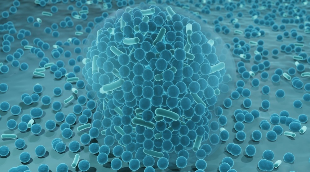
A biofilm is a thin film formed on a material surface or other interfaces due to bacterial activity. A biofilm comprises bacteria, extracellular polymeric substances (EPS), and 80% water. EPS contains nucleic acid, lipids, proteins, and polysaccharides. The biofilm formation is primarily affected by the materials’ surfaces.
The Importance of Proper Biofilm Evaluation Methods for Practical Applications
The properties of bacteria in biofilms vary from those of common airborne bacteria. Biofilms have caused problems in several fields, including materials science, pharmaceuticals, chemical engineering, mechanics, architecture, environmental science, and medicine.
Proper evaluation of biofilms is necessary to control them and develop anti-biofilm materials. Several analytical instruments, such as confocal laser microscopes and optical microscopes, or analytical methods, such as Raman spectroscopy, can be used to qualitatively evaluate biofilms.
Biological methods, including staining methods and gene analyses, can also be used to evaluate biofilms. However, these methods are not suitable for analyzing biofilms for practical applications.
Simple and faster evaluation methods are required that can enable general users, engineers, and researchers to qualitatively and quantitatively evaluate biofilms and directly analyze biofilms over products with unsteady and large shapes.
Crystal violet staining can be used for biofilm evaluation. A color meter is used to analyze different staining situations to determine the biofilm quantity. However, using a color meter for color analyses is expensive and complicated for practical applications. A direct evaluation of the stained specimens is necessary to simplify the evaluation process.
New Method Based on Crystal Violet Staining and Optical Reflection for Biofilm Evaluation
Researchers proposed a new method based on crystal violet staining and optical reflection for quantitative analyses of biofilms. They quantified regular photos of stained specimens using digitized cameras.
Pure titanium and commercially available polyethylene sheet (PE) specimens were used as substrates to verify the proposed method's feasibility for both polymeric and metallic materials. 0.5-1.0 millimeters thick sheets of titanium and PE were cut into 10 × 10 square millimeter pieces and cleaned using alcohol. Escherichia coli (E. coli) were selected as model bacteria as they pose low risk, can be handled quickly and were used frequently in earlier studies.
Biofilm formation was achieved using a static method. Luria–Bertani (LB) liquid medium was autoclaved for 15 minutes at 121 degrees Celsius and then E. coli were added to the medium. The colony formation unit per milliliter was 1 × 10⁹ after a shaking incubation for 24 hours at 37 degrees Celsius.
The bacterial solution was poured into 12 plastic wells, with each well containing 1.2 milliliters of the bacterial solution. The PE and titanium samples were immersed in wells for three, one, and zero days at 25 degrees Celsius in an incubator.
Raman spectroscopy was performed using a Raman spectrometer to confirm the biofilm formation. 0.1% crystal violet aqueous solution was prepared to stain the titanium and PE samples for color analyses.
The extent of biofilm formation on PE and titanium specimens was evaluated through the quantitative analysis of the staining. Optical reflection measured the stained violet color on specimens using color meters. Subsequently, three color parameters, including lightness (L*), red/green value (a*), and blue/yellow value (b*), were combined to obtain the L*a*b* color plane.
The stained specimens' color reflection was analyzed using photo image analyses. A ring light source with a white light emitting diode, a digital camera, and a black box was set up to photograph the specimens.
ImageJ was used to analyze the photographed specimens and obtain histograms of every red, green, and blue (RGB) color on the image. The histograms were then converted into an XYZ color system. The histograms were also converted into the Yxy color system and plotted on the xy chromaticity diagram.
Results obtained using the proposed biofilm evaluation method were compared with those obtained using the colorimetric method.
Significance of the Study
Raman spectroscopy confirmed the formation of biofilms on both PE and titanium specimens, respectively. The comparison of color evaluation results showed that the mean values of image analyses and colorimetric measurements differed.
However, the trend and standard deviation of the color change in the immersed titanium and PE specimens were similar in both methods, which indicated the effectiveness of the image analysis method for biofilm evaluation.
The findings of this study demonstrated that image analyses from photos of specimens stained using crystal violet staining solution could be a suitable alternative to the colorimetric method to quantitatively evaluate biofilms for practical applications.
Reference
Kato, T., Yoshitake, M., Barry, D.M. et al. (2022) Quantitative Analyses of Biofilm by Using Crystal Violet Staining and Optical Reflection. Materials. https://www.mdpi.com/1996-1944/15/19/6727
Disclaimer: The views expressed here are those of the author expressed in their private capacity and do not necessarily represent the views of AZoM.com Limited T/A AZoNetwork the owner and operator of this website. This disclaimer forms part of the Terms and conditions of use of this website.