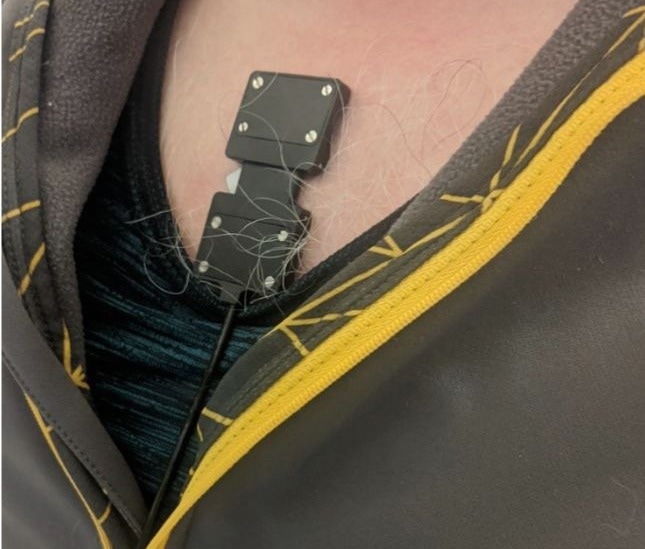
A recent study published in Sensors determines the possibility of gathering respiratory signals from a wearable near-infrared spectroscopy (NIRS) sensor. Researchers used wearable NIRs to differentiate between realistic diseased breathing and normal breathing. NIRS data were examined using a machine learning algorithm to distinguish between realistic diseased and normal breathing.

NIRS sensor affixed over the sternal manubrium. Study: Optical Monitoring of Breathing Patterns and Tissue Oxygenation: A Potential Application in COVID-19 Screening and Monitoring. Image Credit: Mah. A. et al.
Limitations of Current Respiratory Diseases Monitoring Techniques
Techniques for identification, screening, and monitoring of respiratory disease have come under the limelight as a result of the recent outbreak of coronavirus disease (COVID-19). The lack of a quick and reliable screening method was the major problem at the start of the COVID-19 pandemic.
Reverse transcriptase-polymerase chain reaction assays (RT-PCR) are the most frequently used technique for determining COVID-19 infection. The turnaround time for RT-PCR tests can take up to three days in specific cases of mass infection and usually takes hours to confirm results. False-negative test findings are also present, which reduces test sensitivity.
Antigen-detection rapid diagnostic tests (Ag-RDTs) can be used as an alternative to RT-PCR testing to identify COVID-19 active infection. However, these tests frequently produce false-negative results and are less precise than molecular assays. These tests are not monitoring methods; they just reveal the presence of the virus. Ag-RDTs cannot reveal the state of health of the infected individuals. A quick, non-invasive, and affordable method for periodic routine screening and early identification of COVID-19 is needed to stop the spread of infection in high-risk patients.
Monitoring of SpO2 Levels by Pulse Oximetry for Detecting COVID-19
Viral pneumonia caused by COVID-19 results in oxygen deprivation known as silent hypoxia. Tissue oxygen levels begin to decline silently with no other apparent indications. Patients progressively start to breathe more quickly to make up for the hypoxia. Hyperventilation and tissue hypoxia result from this, and the arterial oxygen saturation (SpO2) level declines, leading to hypoxemia.
Standard pulse oximeters (SpO2 90%) are typically used to detect hypoxemia, which causes the patient to breathe more deeply and forcefully, triggering the lung tissue to become more inflamed and damaged in COVID-19. This results in a severe stage of lung injury. Pulse oximetry is not the most reliable approach for early identification of COVID-19 due to technical and physiological limitations, such as limited accuracy.
Acute pneumonia patients also have irregular respiratory patterns that appear shortly after the infection begins. The breathing pattern includes the effort, rhythm, and rate of breathing. Tachypnoea, a cough accompanied by quick and shallow breathing, is a common early symptom of severe pneumonia. As pneumonia worsens, the lungs inflame and inflammatory exudate and sputum fill the alveolar spaces. Hyperventilation is a crucial indicator of severely injured lung tissue. Patients experience low-frequency and shallow breathing when their respiratory centers become depressed, which can be severe and life-threatening if not treated.
Benefits of Continuous Monitoring of Pulmonary Function in COVID-19 Patients
There are several advantages of a precise and dependable approach for tracking pulmonary function in individuals with acute pneumonia. These include categorizing infected individuals according to the severity of their illness, keeping track of how the condition is progressing and how well it is responding to therapy, and creating deadlines for hospital discharge. A precise and non-invasive approach that detects changes in the respiratory pattern can be used for screening, early diagnosis, and follow-up of patients with acute pneumonia, including COVID-19.
The long-term effects of COVID-19 are misunderstood. There is evidence that certain COVID-19-infected people can experience long-term respiratory problems. Therefore, long-term surveillance of lung health and function after infection can give valuable information about the remission or advancement of the disease in the chronic stage. A method for continuously monitoring respiratory function offers advantages compared to clinical tests such as chest radiography, spirometry, and scans that demand trained personnel, as well as trips to specialized facilities.
Potential of Near-Infrared Spectroscopy (NIRS) for Continuous Monitoring of COVID-19
The optical technique known as near-infrared spectroscopy (NIRS) uses light in the near-infrared region of the spectrum (650–1000 nm) to continuously monitor the oxygenation and hemodynamics of tissue.
NIRS offers real-time information regarding tissue oxygenation status by monitoring the variation in concentration of tissue oxygenated (O2Hb) and deoxygenated hemoglobin (HHb) chromophores.
Numerous clinical applications in trauma, urology, neurology, musculoskeletal medicine, and sports medicine have demonstrated the significance of NIRS as a monitoring tool. Several researchers have applied this method to assess respiratory muscle metabolism in sickness.
Non-invasiveness and capacity for real-time monitoring of NIRS can monitor respiratory infectious disorders such as COVID-19. The emergence of hypoxia and hypoxemia, which interfere with the body's oxygenation, is a characteristic of people with acute respiratory diseases. Extraction of respiratory and cardiac characteristics, breathing depth, breathing rate, and heart rate is possible by transcutaneously evaluating NIRS signals from the chest. NIRS offers the ability to identify and track aberrant breathing patterns, decreased tissue, and systemic oxygenation observed in people with respiratory diseases.
Development of Multi-Modal Biosensor with NIRS sensor for Monitoring COVID-19
Mah et al. developed a multi-modal biosensor with a NIRS sensor for diagnosing, screening, and monitoring infectious respiratory disorders. The researchers investigated the viability and effectiveness of utilizing a wearable NIRS sensor to gather respiratory data and differentiate between realistic diseased and normal breathing. A machine learning model was used to differentiate between authentic diseased and normal breathing.
Research Findings
Wearable NIRS sensors can continuously and non-invasively track breathing patterns. The researchers identified three respiratory parameters that differentiate between normal and simulated pathological breathing. The machine learning model distinguished between typical and simulated diseased breathing with a weighted accuracy of 0.87 when all three respiratory parameters were used.
NIRS can potentially be used as a device for monitoring respiratory function and rehabilitation and helping diagnose respiratory diseases by keeping an eye on respiratory function.
Reference
Mah, A. J., Nguyen, T., Ghazi Zadeh, L., Shadgan, A., Khaksari, K., Nourizadeh, M., Zaidi, A., Park, S., Gandjbakhche, A. H., & Shadgan, B. (2022). Optical Monitoring of Breathing Patterns and Tissue Oxygenation: A Potential Application in COVID-19 Screening and Monitoring. Sensors, 22(19), 7274. https://www.mdpi.com/1424-8220/22/19/7274/htm
Disclaimer: The views expressed here are those of the author expressed in their private capacity and do not necessarily represent the views of AZoM.com Limited T/A AZoNetwork the owner and operator of this website. This disclaimer forms part of the Terms and conditions of use of this website.