Jun 14 2021
A novel imaging method can potentially detect neurological diseases, like Alzheimer’s disease, in their earliest stages, thus allowing doctors to diagnose and treat patients more rapidly.
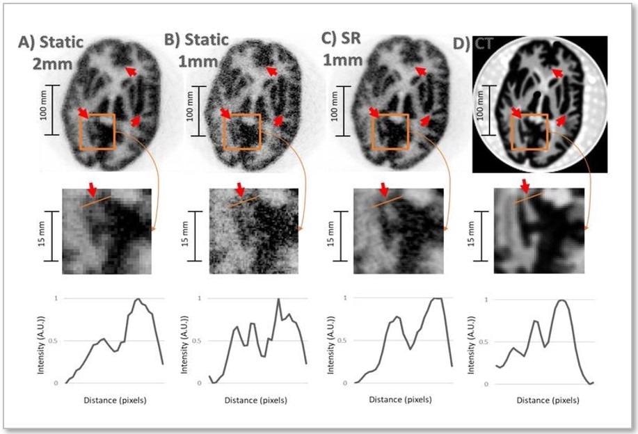
 Result of the Hoffman brain phantom study. Top row: same PET slice reconstructed with (A) 2-mm static OSEM, (B) 1-mm static OSEM, (C) proposed SR method, and (D) corresponding CT slice (note that the CT image can be treated as a high-resolution reference). Middle row: zoom on the region of interest for corresponding images. Bottom row: Line profiles for corresponding data. Image Credit: Society of Nuclear Medicine and Molecular Imaging.
Known as super-resolution, the imaging technique produces incredibly detailed pictures of the brain by integrating positron emission tomography (PET) with an external motion tracking system. This study was presented at the 2021 Virtual Annual Meeting of the Society of Nuclear Medicine and Molecular Imaging.
The quality of images in brain PET imaging is generally restricted by the patient’s unwanted movements at the time of scanning. In this analysis, researchers used super-resolution to harness the subjects’ typically undesirable head movements to improve the resolution in brain PET.
Experiments using moving phantoms and non-human primates were carried out on a PET scanner in combination with an external motion tracking device that constantly quantified the head movement with exceptional precision.
The researchers also performed static reference PET acquisitions without inducing a movement. After combining the data from the imaging equipment, the team was able to recover PET images with visibly greater resolution than that obtained in the static reference scans.
This work shows that one can obtain PET images with a resolution that outperforms the scanner’s resolution by making use, counterintuitively perhaps, of usually undesired patient motion. Our technique not only compensates for the negative effects of head motion on PET image quality, but it also leverages the increased sampling information associated with imaging of moving targets to enhance the effective PET resolution.
Yanis Chemli, MSc, PhD Candidate, Gordon Center for Medical Imaging in Boston, Massachusetts
Although this super-resolution approach has only been checked in preclinical experiments, scientists are presently working to expand it to human beings. Looking ahead, Chemli observed the potentially significant impact of super-resolution on brain diseases, particularly Alzheimer’s disease.
Alzheimer’s disease is characterized by the presence of tangles composed of tau protein. These tangles start accumulating very early on in Alzheimer’s disease—sometimes decades before symptoms—in very small regions of the brain. The better we can image these small structures in the brain, the earlier we may be able to diagnose and, perhaps in the future, treat Alzheimer’s disease.
Yanis Chemli, MSc, PhD Candidate, Gordon Center for Medical Imaging in Boston, Massachusetts