May 17 2021
A new study by Rijksmuseum Boerhaave Leiden and TU Delft demonstrates that a microscope utilized by Antoni van Leeuwenhoek to carry out pioneering research includes a surprisingly ordinary lens.
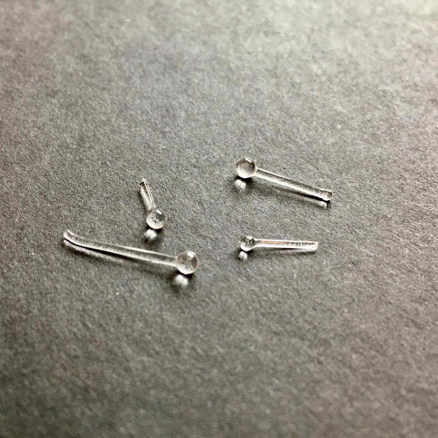
 Microscope lenses reconstructed according to the method of Robert Hooke, which Antoni van Leeuwenhoek also used for his highly magnifying microscopes. Image Credit: Rijksmuseum Boerhaave/TU Delft.
It is a noteworthy finding since Van Leeuwenhoek (1632–1723) guided other researchers to think that his instruments were exceptional. As a result, there have been various guesses regarding his technique for making lenses for over 300 years. The study findings were published in the journal Science Advances on May 14th, 2021.
Early research performed in 2018 has already denoted that a few of Van Leeuwenhoek’s microscopes consisted of general ground lenses. At present, scientists have analyzed a particularly highly magnifying specimen, from the University Museum Utrecht collection. The big surprise in the specimen was that the lens-making technique utilized was a common one, despite having a different type of lens.
Pioneering but Secretive
With the help of his microscopes, Antoni van Leeuwenhoek was able to see a completely new world filled with minute life that no one had ever suspected could survive.
Leeuwenhoek was the first person to visualize unicellular organisms, which is the reason why he is called the father of microbiology. The detail of his visualizations was unparalleled and became outdated only about a century after his death.
Contemporaries of Leeuwenhoek were highly inquisitive regarding the lenses with which Van Leeuwenhoek achieved such amazing feats. But Van Leeuwenhoek was highly secretive about it, indicating that he had discovered a new way of making lenses.
At present, it has been proved to be an empty boast, at least when it comes to the Utrecht lens. This became evident when scientists from Rijksmuseum Boerhaave Leiden and TU Delft subjected the Utrecht microscope to neutron tomography.
It allowed them to analyze the lens without opening the useful microscope and ruining it in the process. The instrument was positioned in a neutron beam at the Reactor Institute Delft, thereby providing a three-dimensional image of the lens.
Small Globule
This lens became a small globule, and the look was similar to a well-known production technique employed in Van Leeuwenhoek’s time. The lens was likely made by placing a thin glass rod in the fire so that the end rolled up into a small ball, which was further broken off the glass rod.
This technique was explained in 1678 by Robert Hooke, another influential English microscopist, which motivated other researchers to follow his path. Also, Van Leeuwenhoek might have taken his lead from Hooke. The latest breakthrough is ironic because it was indeed Hooke who was too inquisitive to learn more regarding Van Leeuwenhoek’s “secret” technique.
The new study illustrates that Van Leeuwenhoek achieved remarkable findings with remarkably ordinary lens production techniques.