Aug 13 2020
Optical-resolution photoacoustic microscopy (OR-PAM) is known to be a robust imaging tool with an excellent potential to image rich optical absorption contrast in biological tissues.
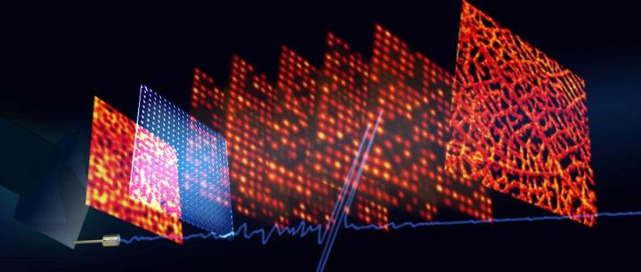
 Image Credit: Yang Li, Terence T. W. Wong, Junhui Shi, Hsun-Chia Hsu, and Lihong V. Wang.
Currently, a majority of the OR-PAM systems depend on mechanical scanning to develop an image, restricting their imaging speed. To address this problem, multifocal OR-PA computed tomography (MFOR-PACT) had been designed by using a microlens array with several optical foci and an ultrasonic transducer array to simultaneously detect PA signals.
But conventional MFOR-PACT systems are complicated and expensive they use an ultrasonic array and the related multi-channel data acquisition system.
In a new study reported in the Light: Science & Applications journal, a research group under the guidance of Professor Lihong Wang from Caltech Optical Imaging Laboratory (COIL), Andrew & Peggy Cherng Department of Medical Engineering and Department of Electrical Engineering, California Institute of Technology, Pasadena, USA, has designed a two-dimensional (2D) MFOR-PAM system using a 2D microlens array for optical excitation and an acoustic ergodic relay for concurrent detection of the PA responses to the multifocal optical illuminations using a single-element ultrasonic transducer.
Dubbed multifocal optical-resolution photoacoustic microscopy through an ergodic relay (MFOR-PAMER), this system can reduce the scanning time by a minimum of 400 times as opposed to traditional OR-PAM systems at the same imaging resolution, while retaining a cost-effective and easy configuration.
This innovative MFOR-PAM system is based on a major enabling element called the acoustic ergodic relay (ER). In the case of PA imaging, an ER—for example, a light-transparent prism—can be employed as an encoder to convert PA signals from various input positions into exclusive temporal signals.
The system impulse response of every input position can be recorded in advance to concurrently detect the PA signals from the complete field-of-view using a single laser shot. Further, mathematical decoding of the encoded PA signals can be performed to rebuild a 2D projection image of the object.
Moreover, the system focuses a wide-field laser beam into several optical focal spots by using a microlens array. In contrast to a traditional focusing lens that requires scanning of a single optical focal spot throughout the entire FOV, the microlens array can decrease the time needed to develop an image by scanning several optical focal spots at the same time.
“Since the excitation pattern through the microlens array is known, each optical focal spot can be computationally localized. By combining the microlens array with the ergodic relay, we can improve the acoustically defined image resolution of the system to the optically defined image resolution and improve the imaging speed by a factor equal to the number of microlens elements.
Researchers of the Study
“Our MFOR-PAMER system has promising potential for many biomedical applications, such as utilizing ultra-violet (UV) illumination for high-speed, label-free histological study of biological tissues. This design can reduce the imaging time from several hours (with a conventional UV OR-PAM system) to less than a minute, significantly improving the efficiency of clinical histology and diagnostics,” the researchers predict.
Journal Reference
Li, Y., et al. (2020) Multifocal photoacoustic microscopy using a single-element ultrasonic transducer through an ergodic relay. Light: Science & Applications. doi.org/10.1038/s41377-020-00372-x.