May 6 2020
Researchers have captured highly detailed images of neurons developed in the lab using extreme ultraviolet radiation, with the potential to help study neurodegenerative diseases.
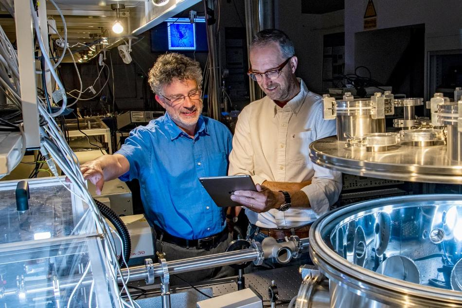
 Professor Jeremy Frey (left) and Dr Bill Brocklesby (right) are pursuing an aim of single molecule imaging. Image Credit: University of Southampton.
Under the guidance of Dr Bill Brocklesby and Professor Jeremy Frey from the University of Southampton, an international study was carried out, in which coherent extreme ultraviolet (EUV) light from an ultrafast laser was used to produce images of the samples by collecting scattered light, without requiring a lens.
The method produced remarkable detail than conventional light microscope images, thereby increasing the chances of possible applications in medicine, such as the study of Alzheimer’s disease.
The study outcomes have been published by the researchers in the Science Advances journal.
The researchers carried out their study at Southampton and at the Artemis facility in the Rutherford Appleton Laboratory, Harwell. The small-scale illustration shows that additional detail can be sampled without the need for huge, high-cost facilities like free-electron lasers and synchrotrons.
The ability to take detailed images of delicate biological structures like neurons without causing damage is very exciting, and to do it in the lab without using synchrotrons or other national facilities is a real innovation.
Dr Bill Brocklesby, Zepler Institute for Photonics and Nanoelectronics, University of Southampton
Brocklesby continued, “Our way of imaging fills an important niche between imaging with light, which doesn't provide the fine details we see, and things like electron microscopy, which require cryogenic cooling and careful sample preparation.”
The collaborative study integrated Southampton’s expertise with Dr Richard Chapman and his colleagues at the Central Laser Facility and included research partners from Italy and Germany.
The EUV imaging method involves using a computer algorithm to process various scatter patterns from a sample. In the project, EUV images of lab-grown neurons from mice were compared with conventional light microscope images, thereby unraveling its highly intricate details. In contrast to hard X-ray microscopy, the delicate neuron structure was not damaged.
It has been a long and sustained effort but highly rewarding. In April 2003, we started a journey with the award of an Engineering and Physical Sciences Research Council Basic Technology grant for New Technology for nanoscale X-ray sources: Towards single isolated molecule scattering.
Jeremy Frey, Professor and Head of Computational Systems Chemistry, University of Southampton
Frey continued, “Some 17 years later, almost to the day, our paper in Science Advances demonstrates that the effort was well worth the hard work of our interdisciplinary team, obtaining the first ultra-high resolution images of a real biological sample using coherent soft-x-ray microscopy (ptyography).”
We are looking forward to applying our microscope to many biological, chemical and material problems. We continue to pursue even higher resolution with the ultimate aim of singe molecule imaging, a goal that now seems very much in view.
Jeremy Frey, Professor and Head of Computational Systems Chemistry, University of Southampton
Although EUV microscopy offers several benefits over hard X-ray, optical, or electron-based methods, to date, conventional EUV optics and sources have incurred huge associated costs and scale.
The focus of the new method is on nonlinear optical techniques and, specifically, from high harmonic generation (HHG) with the help of strong femtosecond lasers. Using the study outcomes, Oxford’s Artemis team is working toward achieving regular access to this method in the upcoming days.
Moreover, the combination of tomographic imaging methods and such new advancements in laser technologies and coherent EUV sources seems promising for high-resolution biological imaging in 3D.
Journal Reference:
Baksh, P. D., et al. (2020) Quantitative and correlative extreme ultraviolet coherent imaging of mouse hippocampal neurons at high resolution. Science Advances. doi.org/10.1126/sciadv.aaz3025.