Mar 9 2018
Similar to photography, holography is a means to record everything around us. Both technologies involve the use of light to perform recordings; however, in the place of two-dimensional images, holograms reproduce three-dimensional shapes.
The shape can be figured out from the patterns formed when light is bounced off an object and interferes with another light wave that acts as a reference.
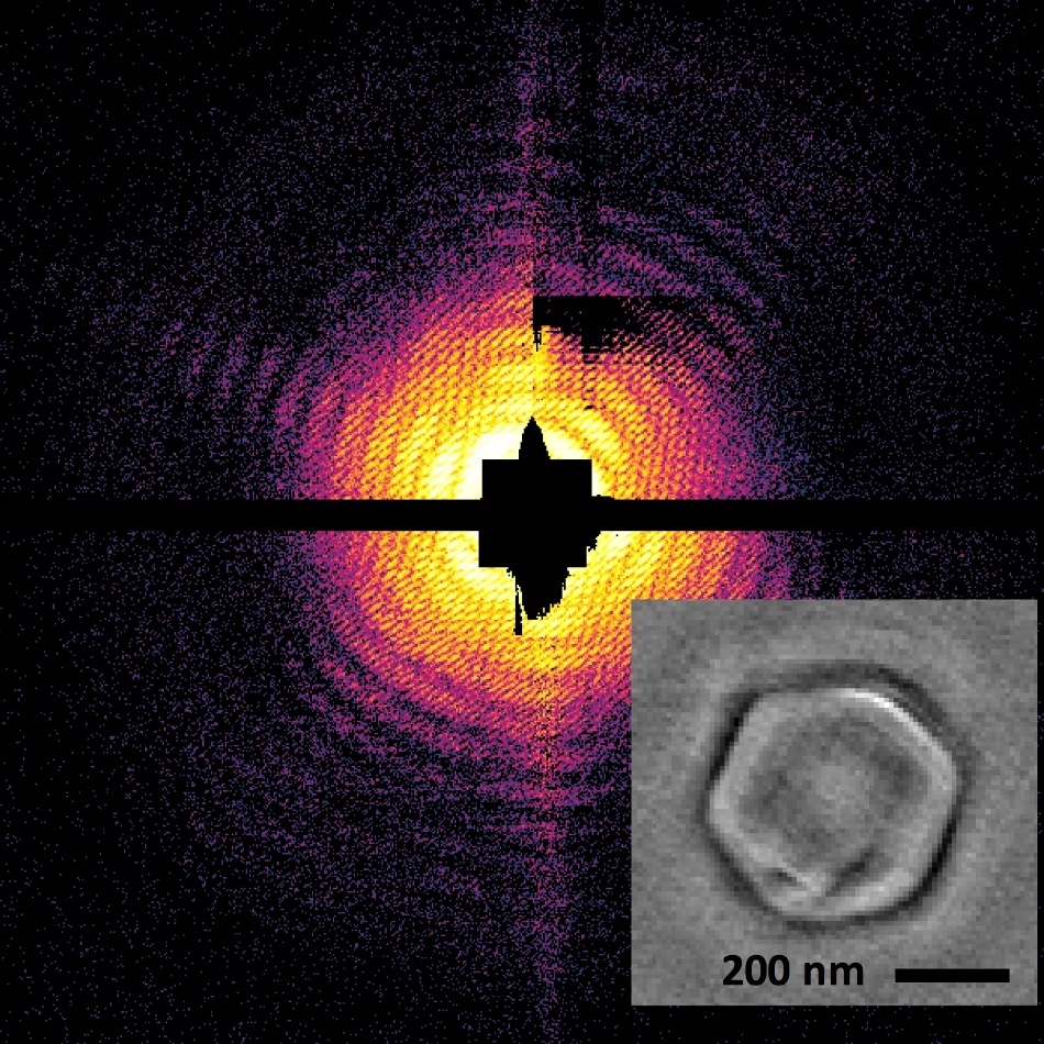
 Image credit: Anatoli Ulmer and Tais Gorkhover/The Technical University of Berlin and SLAC National Accelerator Laboratory.
Upon being developed by using X-ray light, holography could be a significantly helpful technique for acquiring high-resolution images of a nanoscale article, which is very small, and its size is evaluated in nanometers or one-billionths of 1 m.
To date, X-ray holography has been limited to articles that form crystals or has been dependent on the cautious positioning of the sample on a surface. Yet, several nano-sized particles are short-lived, non-crystalline, and very brittle. They might even be impaired by damage or changes at the time of an experiment while being positioned on a surface. Exotic states of matter, aerosols, and the smallest life forms normally belong to these classes and hence are challenging to analyze by using traditional imaging techniques.
As part of the latest work featuring on the cover of Nature Photonics in March 2018, scientists have devised an innovative holographic technique known as in-flight holography. By using this technique, the researchers could demonstrate the first ever X-ray holograms of nanoscale viruses that were not fixed to any surface.
The patterns required to develop the images were obtained from the Linac Coherent Light Source (LCLS), the X-ray free-electron laser at the Department of Energy’s SLAC National Accelerator Laboratory. Nanoviruses have been investigated at LCLS in the absence of a holographic reference, yet the interpretation of the X-ray images mandated several steps, was dependent on human input and was a computationally difficult procedure.
In the new research, X-ray light scattered from the virus was superimposed with X-ray light scattered from a reference nano-sized sphere. The curvature included in the superimposed images from both the objects offered in-depth information and details related to the shape of the mimivirus, a virus with a width of 450 nm. This procedure remarkably simplified the data interpretation.
“Instead of thousands of steps and algorithms that potentially don’t match up, you have a two-step procedure where you clearly get the structure out of your image,” stated Tais Gorkhover, lead author of the study, who is a Panofsky Fellow at SLAC and researcher at the Stanford PULSE Institute.
At present, the researchers can perform their reconstruction of a sample within fractions of a second or even quicker by using the holographic technique.
Before our study, the interpretation of the X-ray images was very complicated and the structure of nanosamples was reconstructed long after the actual experiment using non-trivial algorithms. With ‘in-flight’ holography, the procedure is very simple and in principle can be performed while taking data. This is a real breakthrough.
Christoph Bostedt, Co-Author & Scientist - DOE’s Argonne National Laboratory
“Another advantage of the in-flight holography method is that it is less prone to noise and to the artifacts that can appear in the detector compared to non-holographic X-ray imaging,” stated Anatoli Ulmer, a co-author and Ph.D. student from the Technical University of Berlin in Germany.
The scientists propose that in the future, in-flight holography will prove to be an innovative means to analyze combustion, air pollution, and catalytic processes, where nanoparticles are involved in all these processes.
An international group of researchers from the DOE’s Lawrence Berkeley and Brookhaven national laboratories, the Chalmers University of Technology in Sweden, the Czech Academy of Science, Deutsches Elektronen-Synchrotron (DESY) in Germany, the Institute of Optics and Atomic Physics in Germany, the Institute for Solid State Physics and Optics in Hungary, KTH Royal Institute of Technology in Sweden, Northwestern University, and Uppsala University in Sweden also contributed to this study.
LCLS is a DOE Office of Science user facility.
Public Lecture | Holograms at the Nanoscale: New Imaging for Nature's Tiniest Structures
Watch Tais Gorkhover, a Panofsky Fellow at SLAC and researcher at the Stanford PULSE Institute, explain the holographic method in detail in her recent public lecture, “Holograms at the Nanoscale: New Imaging for Nature's Tiniest Structures.” (Video credit: SLAC National Accelerator Laboratory)