Jan 26 2018
Detecting a number of diseases via PETs, X-rays, MRIs or CAT scans is today considered to be regular practice. However, all of these medical imaging methods come with some risk of radiation and take long hours – if not days – to obtain results. Most prominently, the level of information they offer is lacking, since it is not at the molecular level.
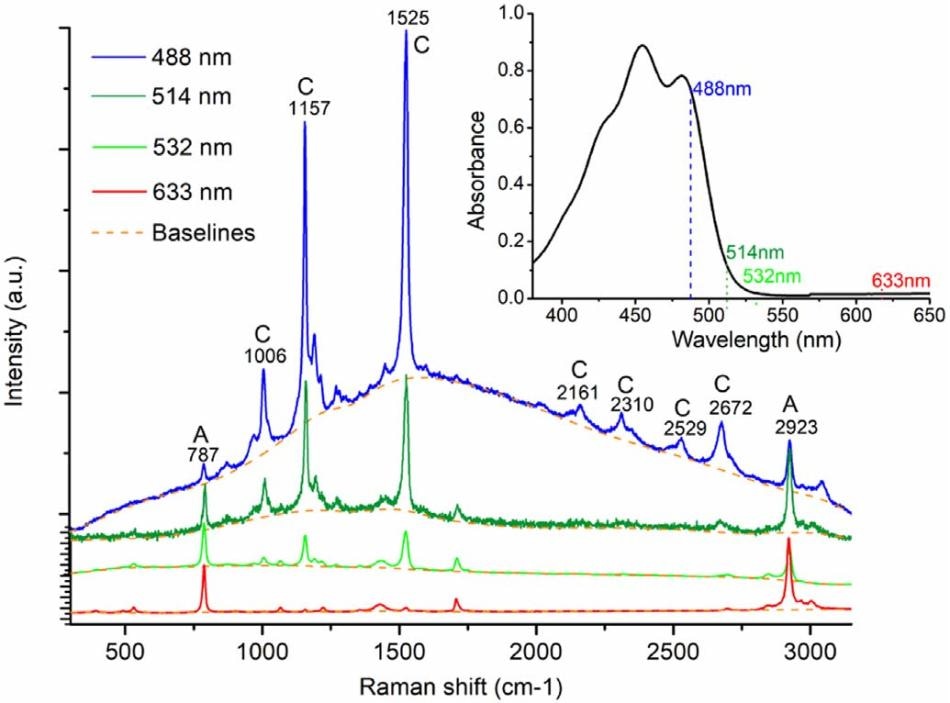
 Raman spectra of beta-carotene solution excited by visible laser beams. (Image credit: The City College of New York)
Raman spectroscopy, a tool previously employed for providing molecular information in science, is presently being used in biomedicine to provide an optical biopsy that provides more in depth, faster detection. This innovative, less invasive method of detecting disease employing light salient properties was used for the very first time in 1991 in order to identify a fingerprint for cancer in tissue, by a team headed by Robert Alfano, a renowned Professor of Science and Engineering at The City College of New York, and director of The Institute for Ultrafast Spectroscopy and Lasers (IUSL) of the City University of New York at City College.
A recent IUSL paper featured in the Journal of Photochemistry and Photobiology present a report on how research in this area continues to improve. It demonstrates how Resonance Raman spectroscopy in tissue (specially investigating carotene using varied visible lasers) can identify vibrations when an exciting laser makes an entry into an absorption of a molecule. The paper’s authors are visiting scientists Luyao Lua of Wenzhou Medical University, and Lingyan Shi of Columbia University, along with City College IUSL Research Associate Jeff Secor, and Robert Alfano.
Resonant Raman using the laser pointer 532 nm has become an efficient tool for investigating molecular components in tissues and cells, providing more detailed information and a way to detect diseases like skin cancer, brain cancer, or atherosclerosis – in mere seconds.
Robert Alfano