May 26 2017
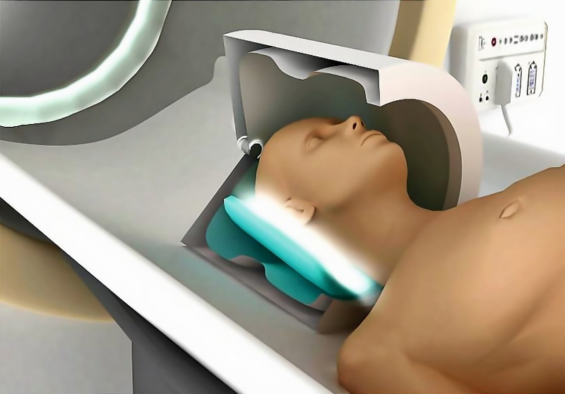
 A patient in MRI scan modified by metasurface. CREDIT Copyright ITMO University
A new metasurface-based technology has been designed and tested by scientists from the Netherlands and Russia in order to improve the local sensitivity of MRI scanners on human test subjects for the very first time.
The metasurface is made up of strips that are periodically arranged. Placed under a patient's head, the metasurface offers a superior image quality from the local brain region. The results featuring in Scientific Reports demonstrates that the use of metasurfaces can potentially decrease image acquisition time, enable the procedure to become more comfortable for patients and attain images with higher resolution in order to allow diseases to be diagnosed at an earlier stage.
Magnetic resonance imaging (MRI) is a medical technology that has been extensively used for the examination of internal organs, which can provide, for instance, information on functional and structural damage in cardiovascular, neurological and musculoskeletal conditions. MRI also plays a vital role in oncology. However, acquiring an MRI scan takes much longer than an ultrasound scan or computed tomography due to its intrinsically lower signal-to-noise ratio. This explains that a patient will have to lie motionless within a confined apparatus for almost one hour, leading to major patient discomfort, and comparatively long lines in hospitals.
For the very first time, specialists from Leiden University Medical Center in the Netherlands and ITMO University have attained human MR-images with improved local sensitivity provided by a thin metasurface, which refers to a periodic structure of conducting copper strips. These elements were attached to a thin flexible substrate and then incorporated into the close-fitting receiving coil arrays inside the MRI scanner.
We placed such a metasurface under the patient's head, which increased local sensitivity by 50%. This allowed us to obtain detailed scans of the occipital cortex in half the usual time. Such devices could potentially reduce the duration of MRI studies and improve its comfort for subjects.
Rita Schmidt, Researcher, Department of Radiology, Leiden University Medical Center
The metasurface, positioned between the receive coils and a patient, improves the signal-to-noise ratio in the region of interest.
This ratio limits the MRI sensitivity and duration of the procedure. Often the scans must be repeated many times and the signals added together to separate actual data from random noise. The use of metasurfaces eliminates this requirement. If now an examination takes twenty minutes, it may only take ten in the future. If today’s hospitals serve ten patients a day, they will be able to serve twenty using our development.
Alexey Slobozhanyuk, Research Fellow, International Laboratory of Applied Radioengineering, ITMO University
The scientists believe that the metasurface is capable of increasing the definition of the resulting scans.
"The size of voxels, or 3D-pixels, is also limited by the signal-to-noise ratio. Instead of accelerating the procedure, we can adopt an alternative approach and acquire more detailed images in the same amount of time as before. Such images can let us detect tumors at earlier stages", says Andrew Webb, leader of the project and professor of Radiology at Leiden University Medical Center.
Till date, no one has succeeded in combining metamaterials into close-fitting receiver arrays as the dimensions of the metasurfaces were extremely wide. This problem was solved by the novel 8 mm-wide design of the metasurface.
Our technology can be applied for production of metamaterial-based ultra-thin devices for many different types of MRI scans, but in each case, one should firstly carry out a series of computer simulations as we have done in this project. One needs to make sure that the MRI device’s properties do not affect metasurface’s performance.
Rita Schmidt, Researcher, Department of Radiology, Leiden University Medical Center
The scientists are now planning to continue their research in both St. Petersburg and Leiden, and they are also hoping to transform ITMO University’s Laboratory of Applied Radioengineering into a center for cutting-edge projects in this field in order to attain modern equipment and motivate more young specialists to develop prototypes and launch them into clinical practice. For that reason, a grant request has already been filed at Russian Science Foundation by the staff at Applied Radioengineering Laboratory and Andrew Webb.