Dec 14 2015
Bluetongue disease is a viral infection that has killed approximately 2 million cattle in Europe over the past two decades.
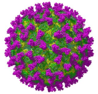
 Cryo-electron microscope image of a Bluetongue virus.
A new study has revealed the atomic structure of the Bluetongue virus, including the means by which it infects healthy host cells. Scientists hope to use this information to aid in the creation of vaccines and drug treatments for bluetongue disease.
A team led by Hong Zhou, a professor of microbiology, immunology and molecular genetics and faculty director of the Electron Imaging Center for Nanomachines at UCLA’s California Nanosystems Institute, collaborated on the research with a team led by Polly Roy, professor of virology at the London School of Hygiene and Tropical Medicine. The research was published in the journal Nature Structural and Molecular Biology.
Using cryo-electron microscopy, the researchers discovered the Bluetongue virus’s two-step process for infecting healthy cells.
The virus has sensor proteins on its surface that detect changes in the acidity of its environment. When these proteins sense higher acidity caused by proximity to the target cell, the virus unfurls a protein structure that penetrates the outer membrane of the cell and anchors to it, causing infection.
The scientists confirmed this mechanism by lowering the acidity around the virus, which caused the protein structure to detach and refold.
“The advantage we have with cryo-electron microscopy is its ability to resolve three-dimensional structures of nanoscale objects in their native environments. This ability, with the revolutionary technology for counting electrons, enables us to collect vast data that we use to reconstruct three-dimensional images of these biological structures, often for the first time,” Zhou said.
“We are delighted with these results, which show the virus in the highest possible detail at different pH levels,” Roy said. “This represents a key piece in the puzzle and a significant step forward for understanding molecular structures and mechanisms in this family of viruses. We hope it will enable the design of specific antiviral agents and new and efficient vaccines for the control of bluetongue and related viral infections of animals and humans.”
The study’s lead author is Xing Zhang, scientific director of the Electron Imaging Center for Nanomachines. Other authors are Xuekui Yu, a project scientist in molecular biology, immunology and molecular genetics at UCLA, and Avnish Patel and Cristina C. Celma of the London School of Hygiene and Tropical Medicine.
This research was supported by the National Institutes of Health, the National Science Foundation, UCLA and the Welcome Trust, UK.