Sep 11 2015
The New Jersey Commission on Spinal Cord Research awarded $198,000 over two years to Zhiguo (Tony) Jiang, Ph.D., research scientist at Kessler Foundation, to examine the efficacy of using diffusion tensor imaging (DTI) in assessing recovery after spinal cord injury (SCI). Results may enable medical professionals to predict motor recovery and provide more accurate prognoses.
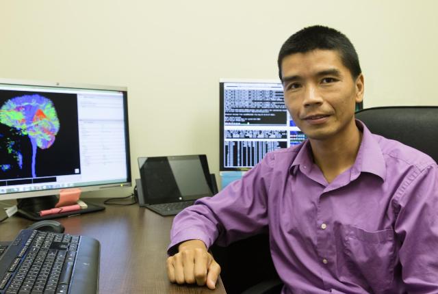
 This is Dr. Jiang sitting in front of two computer screens, which depict a colorful brain scan and data. Credit: Kessler Foundation
DTI, an advanced, non-invasive form of MRI technology, measures the diffusion of water elements in nerve fibers to detect structural changes above and below the level of injury. The study funds 150 MRI scans at the Rocco Ortenzio Neuroimaging Center at Kessler Foundation--the only free-standing imaging center in the U.S. solely dedicated to rehabilitation research.
"In addition to physical challenges, a spinal cord injury can also impose significant financial and emotional burdens on the individual and the family," said John DeLuca, Ph.D., senior vice president of Research and Training and director of the Ortenzio Center. "We are seeing advancements in recovery following rehabilitation, but little objective evidence. Using neuroimaging, we can document how the spinal cord recovers. With this knowledge, we can determine the best course of treatment and provide better assessments of functional recovery so individuals can properly prepare for the months and years ahead."
Individuals with incomplete SCI enrolled in the study will receive standard rehabilitative therapies. DTI measurements will be taken at baseline and one, two, four and six months after the treatment program. It is expected that structural integrity will be compromised in individuals with SCI compared with controls. In addition, integrity of the fiber structure is expected to improve with spontaneous recovery and rehabilitation. Improvement in integrity should correspond with improved mobility in the individual with SCI.
Unlike traditional neurological tests, neuroimaging can assess function below the level of injury. While progress has been made in imaging of the brain, spinal cord imaging remains a new frontier in research and clinical practice. Obtaining detailed pictures of the spinal cord is difficult due to its thin and flexible nature. Researchers at Kessler Foundation are exploring various techniques and imaging methods to advance applications of spinal cord imaging.
There are 12,000 new cases of SCI in the U.S. each year. The average lifetime costs of a single injury can be in the millions.