Nov 4 2014
A new application note from Olympus explores how, moving into the future of cultural heritage research, the latest high-resolution light microscopy is rapidly advancing our knowledge of the past.
Discovering and understanding novel strategies for historic conservation, Dr Olivier Schalm’s research at the Department of Conservation Studies, University of Antwerp (Belgium), has benefited from the latest in high-resolution light microscopy with the Olympus DSX500 Opto-digital system.

Introducing cutting-edge digital imaging technologies and retaining true colour information at magnifications of up to 4,000x, this approach has even presented a fast and efficient alternative to scanning electron microscopy (SEM).
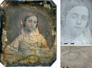
Light microscopy is a popular analytical tool for cultural heritage researchers, gathering valuable information on the degradation of historic artefacts such as antique glass and daguerreotype photographic plates. Only by truly understanding the mechanisms of degradation can one develop and optimise techniques to control and slow the process, or restore an object to a past state. Such aims form the major focus of Dr Schalm’s research efforts.
Investigations often call for greater resolution than possible with standard light microscopy, however, and in these cases Dr Schalm would previously turn to SEM. Unfortunately, high resolution and chemical composition data does come at the cost of colour information and spatial context. In contrast, the Olympus DSX500 retains true-colour information and is capable of reaching 4,000x magnification.
Utilising the latest digital imaging technologies presents a fast, efficient and accessible alternative to SEM, and Dr Schalm found that in the great majority of cases, magnification above 4,000x was unnecessary.
As part of the Olympus Opto-digital series, the DSX500 is designed for materials science and industrial quality control applications. Combining advanced optics with powerful digital capabilities creates a sophisticated yet intuitive system, capturing high-resolution images at the highest quality, in either 2D or 3D.
Product and Application Specialist for Materials Science Microscopy at Olympus, Markus Fabich comments: “Although we designed and developed the Olympus Opto-digital series with industrial quality control in mind, Dr Schalm’s ground-breaking work has demonstrated just how flexible these systems really are, capable of extracting an incredible level of information from a diverse array of samples throughout a broad range of applications”.
For both glass and metallic artefacts, the information revealed was rather unexpected, opening new avenues of exploration into the mechanisms underlying degradation.