Oct 10 2014
Vikram Kodibagkar’s progress in engineering tools and techniques to more precisely reveal the condition of body tissues, specifically tissue oxygen levels, has earned support for his research from the National Science Foundation (NSF).
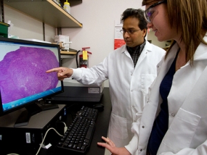
 Vikram Kodibagkar (left), an assistant professor in ASU's School of Biological and Health Systems Engineering, is the recipient of a National Science Foundation CAREER award for his work to more precisely reveal the condition of body tissues, specifically tissue oxygen levels. Photo by: Jessica Hochreiter/ASU
He’s the recipient of an NSF CAREER award, which recognizes emerging education and research leaders in engineering and science. The award provides $440,000 over five years to fund Kodibagkar’s work to expand the capabilities of medical imaging technology. The project will also help provide research and educational opportunities for students.
Kodibagkar is an assistant professor in the School of Biological and Health Systems Engineering, one of the Ira A. Fulton Schools of Engineering at Arizona Sate University. His focus is on developing techniques using magnetic resonance imaging (MRI) to perform oximetry – measuring the amount of oxygen in body tissue.
Hypoxia, or lack of oxygenation – due to impaired delivery of oxygen to tissue and/or increased oxygen consumption by tissue cells – can be a sign of disease or injury, such as cancer, stroke or traumatic brain damage.
“What we are trying to do is figure out how damaged or diseased tissue will change, either by itself or in response to specific therapies,” he said. “And we want to do this early on in the onset of disease through noninvasive prognostic imaging.”
One of Kodibagkar’s research goals is to “develop a theoretical framework for methods that would enable us to extract quantitative information about tissue oxygenation from MRI data,” he said.
“That would allow us, for example, to compare changes that occur in a tumor over time or in response to treatment. You could potentially look at its condition in the early stages of a disease and predict what will happen from that point on. This would give you the basis for planning an effective medical treatment,” he explained.
He’ll pursue this objective by using two novel oximetry techniques he has developed. One technique involves binding a small molecular MRI contrast agent (a chemical compound) that targets specific areas of the body. The other involves examining changes in the MRI properties of a nanoprobe to determine oxygen levels.
He will also work on further developing methods of delivering and distributing the molecular agent and the nanoprobe to targeted areas so they can provide MRI imaging that gives accurate information about oxygenation.
Studies are providing increasing evidence that tumors have high concentrations of hypoxic cells – cells that are inadequately oxygenated. The severity of hypoxia can be a strong factor in predicting the progression of disease, particularly cancer.
The potential effectiveness of chemotherapy, radiotherapy and photodynamic therapy to treat cancer can often be determined by knowing the various levels of concentrations of hypoxic cells. For instance, Kodibagkar said, hypoxia in solid tumors can make those tumors resistant to some anticancer drugs, and the use of a single drug may be ineffective, or worse, leave behind a malignant subpopulation of tumor cells.
He wants to produce more accurate evaluations of tumor oxygenation to provide a better understanding of how particular tumors would respond to different therapeutic combinations, potentially enabling therapy to be tailored to characteristics of the conditions of individual patients.
The NSF CAREER award also will aid Kodibagkar’s efforts to increase involvement of ASU graduate students in lab work related to his research, and to establish a hands-on training program in imaging technologies, to be called HOSPIT, for high school students and undergraduates.
“Powerful imaging tools are emerging that are expanding career options in the field, but there are very few opportunities for younger students to be exposed to this,” Kodibagkar says.
For that training, he plans to use two instruments – a commercial bench-top MRI machine and “shoebox fluorescent imaging system” – the latter designed and constructed by Kodibagkar for a course he co-taught with Karmella Haynes and Sarah Stabenfeldt, fellow assistant professors in the School of Biological and Health Systems Engineering.
His plan is to use the training programs as the basis for developing more extensive courses in medical imaging, including courses that could be offered online. The first trial of HOSPIT training program was successfully conducted this summer with five high school students.
Beyond that, he hopes an online education component will aid efforts to recruit students from around the world to college programs offering advanced education in medical imaging – particularly prospective students in underdeveloped countries where medical imaging expertise is especially needed.