A research team led by Peidong Yang, a chemist at the Materials Sciences Division of the Lawrence Berkeley National Laboratory (Berkeley Lab), has developed a nanowire endoscope by integrating a waveguide of tin oxide nanowire with an optical fiber’s tapered end, paving the way for capturing optical images of the inside of an individual live cell at high resolution or accurately delivering drugs, proteins, genes or other cargo without causing damage to the cell.
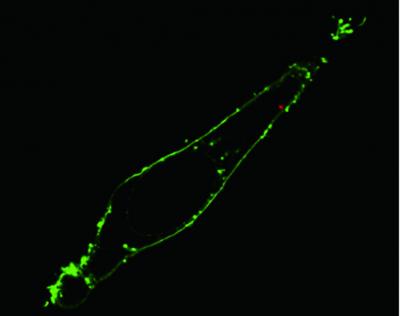
 Fluorescence confocal image of a single living HeLa cell shows that via nanoendoscopy a quantum dot cluster (red dot) has been delivered to the cytoplasm within the membrane (green) of the cell. (Credit: (Courtesy of Berkeley Lab))
This mechanically strong and highly flexible nanowire optical probe also finds applications in single-cell electrophysiology and biosensing. It is possible to couple the light that passes through through the optical fiber with the nanowire from where the light is released into open space subsequent to its arrival at the tip. The nanowire tip has high suppleness thanks to its high aspect ratio and nano-size and can be utilized several times, as it can withstand continual buckling and bending.
Yang stated that based on the benefits of fiber-optic fluorescence imaging and nanowire waveguides, the team was able to control the light inside live cells for performing high temporal and spatial resolution study of biological processes inside the cells. The team also demonstrated that its nanowire endoscope can sense optical signals of sub-cell regions and can deliver temporal and spatial specified cargos into the cells using light-stimulated mechanisms, he added.
Advancements in nanophotonics overcome the diffraction blockade of visible light microscope that prevents its usage in studying objects smaller than one-half of the incident light wavelength, paving the way for using optical imaging systems to study sub-cellular components. However, these optical systems are bulk in size, expensive and complex.
To demonstrate its nanowire endoscope’s capability as a sub-cellular imaging light source, the team coupled its system optically with an excitation laser and subsequently waveguided blue light through the membrane and into the inside of single HeLa cells. Yang stated that the optical output delivered a highly localized and focused illumination as its emission from the endoscope was tightly confined to the tip of the nanowire. Moreover, the introduction of the nanowire did not cause any damage to the cells, he added.
To demonstrate the capability of the system for payload delivery to target sites within a cell, the team utilized photo-activated linkers, which can be sliced by low-power UV radiation, to bond quantum dots to the tip of the nanowire. The functionalized nanowire endoscope delivered the quantum dot payload into the specific intracellular sites within a minute. According to Yang, his nanowire endoscope can be used to optically or electrically excite a live cell in the future.