Nov 10 2010
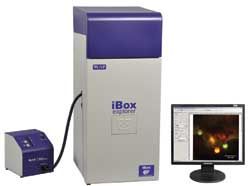
UVP will launch a new iBox Explorer fluorescence imaging microscope at the ‘Neuroscience 2010’ event. The UVP iBox Explorer can micro image organs and cells subcutaneously and can also image the body cavity of live mice.
 iBox Explorer Fluorescence Imaging Microscope
The fluorescence imaging microscope functions through an intuitive software control with parfocal and parcentered optical configurations. As a result, the operation leads to faultless imaging via the magnification ranges.
The iBox Explorer comprises a cooled color camera with a high frame rate and a technology that offers high throughput, image capture and quick detection. The microscope is suitable for imaging micro metastases, vasculature, tumor micro environment, tumor/host interactions and margins, and whole organs.
The UVP iBox Explorer enters the company’s line of complete mouse imagers such as iBox Spectra and iBox Scienta to transform the in vivo fluorescent imaging.