 By Owais AliReviewed by Lexie CornerMar 26 2024
Optical technologies have revolutionized medical diagnostics, surgery, and therapeutics over recent decades. However, their effectiveness relies on intricate light-tissue interactions, which are notably influenced by skin color. This article explores this correlation and its implications for medical optical technologies, highlighting the need for inclusive and equitable healthcare solutions.

Image Credit: Prostock-studio/Shutterstock.com
Optical Technologies in Medical Fields
Optical technologies are driving transformation in medical diagnostics, surgery, and therapeutics, representing a $73 billion global medical market. These technologies, ranging from microscopes and ophthalmoscopes to robotic-assisted imaging systems, have revolutionized visual examination and minimally invasive procedures.1
One of the most widely used optical technologies in medicine is optical coherence tomography (OCT), which employs low-coherence interferometry to generate high-resolution, cross-sectional images of tissues. This is crucial for diagnosing and managing glaucoma and retinal disorders.
Beyond imaging, optical technologies have also transformed therapeutic approaches. Photodynamic therapy (PDT) harnesses the power of light-activated photosensitizers to selectively target and destroy cancerous or diseased cells, offering a minimally invasive treatment option for various malignancies and skin conditions.2
However, the fundamental operation of these various optical devices revolves around their interaction with tissue via light absorption and scattering. Absorption transforms light energy into heat or triggers photochemical reactions, while scattering leads to the broadening and attenuation of light beams as they traverse through tissue.
These fundamental interactions determine how deeply light penetrates to form a focus for measurements or therapy delivery. They also influence the strength of optical signals returning from within tissue, which imaging and diagnostic technologies rely upon.3
Therefore, a comprehensive understanding of these optical device skin interactions is crucial for developing and efficiently using medical optical technologies.
The Critical Role of Skin Color
One of the most significant factors influencing the optical properties of human tissue is skin color, primarily determined by melanin concentration.
Melanin is a highly absorbent chromophore, particularly in the visible and near-infrared (NIR) regions of the electromagnetic spectrum. The concentration of melanin varies widely among individuals due to genetic factors and sun exposure, exerting a substantial impact on its optical properties. Individuals with higher melanin concentrations, resulting in darker skin tones, exhibit greater light absorption and scattering within the skin, affecting light transmission through the tissue.
This variation in absorption and scattering properties due to skin color poses significant challenges for medical optical technologies.1
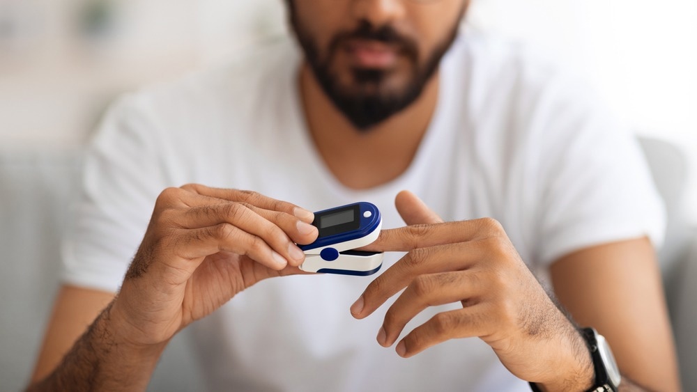
For instance, pulse oximetry, a widely employed non-invasive method for measuring blood oxygen saturation, may encounter accuracy challenges in individuals with darker skin tones. The increased melanin absorption can lead to inaccurate arterial oxygen saturation estimations, potentially delaying hypoxemia detection and impacting patient care.4
Similarly, skin pigmentation can adversely affect image quality and oximetry measurements in photoacoustic imaging (PAI), which utilizes light absorption to generate acoustic signals. Higher melanin concentrations in darker skin tones can lead to increased acoustic clutter and reduced depth penetration, potentially obscuring underlying vascular structures and introducing inaccuracies in oxygen saturation readings.5
Another area where skin color variations pose challenges is in laser treatments and PDT. The efficacy of these treatments relies on the precise delivery of light energy to target tissues. However, variations in melanin concentration can impact the depth of light penetration, potentially leading to suboptimal treatment outcomes or an increased risk of side effects.2
These challenges underscore the importance of considering variations in skin color’s optical properties for inclusive medical device design and application, as failure to do so can result in misdiagnoses, suboptimal treatment outcomes, and potential health disparities among diverse populations.
Insights from Recent Studies and Case Studies
Impact of Skin Color Variations on Photoacoustic Oximetry Measurements
In a study published in the Biomedical Optics Express, UC San Diego researchers explored the influence of skin tone on photoacoustic (PA) oximetry, a potential alternative to pulse oximetry.
Their findings revealed that individuals with darker skin tones exhibited higher PA signal intensity at the skin's surface due to increased melanin content, leading to more acoustic clutter and lower oxygen saturation readings in radial arteries than those with lighter skin tones.
To address this, the researchers proposed corrective techniques, such as time gain compensation adjustments, noise reduction algorithms, and a basic correction factor, which brought PA oximetry measurements to within 2 % of those from pulse oximetry.6
Impact of Melanosome Volume on Transcutaneous Bilirubin Levels
A study published in Pediatric Research assessed the effect of skin color on transcutaneous bilirubin (TcB) measurements, a vital method for diagnosing hyperbilirubinemia in newborns.
The researchers found that higher volume fractions of melanosome (which package melanin), alongside increased bilirubin levels, resulted in larger underestimations of TcB compared to unpigmented skin, with underestimations ranging from 26 to 132 µmol/L at a TcB level of 250 µmol/L.
However, these measurements can be improved by incorporating multi-wavelength measurements and melanin-specific corrections.7
The Future of Medical Optics: Addressing the Impact of Skin Color
Skin color significantly influences the light-tissue interactions that govern medical optics. Understanding these implications is crucial for designing technologies capable of providing effective, safe, and unbiased care, irrespective of patient attributes.
Advanced computational models and machine learning techniques offer promising ways to predict light behavior in tissues with varying melanin concentrations and automatically compensate for skin color effects, enhancing device accuracy. Standardized testing methods, including tissue-mimicking phantoms, will also ensure thorough validation across different skin tones.6
However, interdisciplinary collaborations among optics, dermatology, and biomedical engineering experts are needed to develop holistic solutions that cater to diverse patient populations and promote equitable healthcare access.
More from AZoOptics: Holographic Optical Elements for Imaging and Optical Processing
References and Further Reading
- Setchfield, K., Gorman, A., Simpson, AHR., Somekh, MG., Wright, AJ. (2024). Effect of skin color on optical properties and the implications for medical optical technologies: a review. Journal of Biomedical Optics. doi.org/10.1117/1.JBO.29.1.010901
- Huang, YY., Vecchio, D., Avci, P., Yin, R., Garcia-Diaz, M., Hamblin, MR. (2013). Melanoma resistance to photodynamic therapy: new insights. Biological chemistry. doi.org/10.1515/hsz-2012-0228
- Keiser, G. (2016). Light-tissue interactions. Biophotonics: Concepts to applications. doi.org/10.1007/978-981-10-0945-7_6
- Afshari, A., et al. (2022). Evaluation of the robustness of cerebral oximetry to variations in skin pigmentation using a tissue-simulating phantom. Biomedical Optics Express. doi.org/10.1364/BOE.454020
- Vogt, WC., Wear, KA., Pfefer, TJ. (2023). Phantoms for evaluating the impact of skin pigmentation on photoacoustic imaging and oximetry performance. Biomedical Optics Express. doi.org/10.1364/BOE.501950
- Mantri, Y., Jokerst, JV. (2022). Impact of skin tone on photoacoustic oximetry and tools to minimize bias. Biomedical Optics Express. doi.org/10.1364/BOE.450224
- Dam-Vervloet, AJ., Morsink, CF., Krommendijk, ME., Nijholt, I. M., van Straaten, H. L., Poot, L., Bosschaart, N. (2024). Skin color influences transcutaneous bilirubin measurements: a systematic in vitro evaluation. Pediatric Research. doi.org/10.1038/s41390-024-03081-y
Disclaimer: The views expressed here are those of the author expressed in their private capacity and do not necessarily represent the views of AZoM.com Limited T/A AZoNetwork the owner and operator of this website. This disclaimer forms part of the Terms and conditions of use of this website.