Understanding how biomolecules function and interact is fundamental to biochemistry, and can aid efforts to develop new drugs and medical treatments, but these biomolecules were not compatible with high vacuum conditions and the intense electron beams used in techniques such as transmission electron microscopy.
As early as the 1960s, scientists realised that although transmission electron microscopy (TEM) allowed them to examine the structure of molecules and materials at the atomic scale, the technique was too harsh for delicate biomolecules. In TEM, a beam of electrons passes through a very thin sample and interacts with molecules to yield an image of the sample on a detector. However, the technique is quite damaging, evaporating the water surrounding the molecule, and burning or destroying the molecule with the high energy electron beam.
Scientists considered a variation of TEM, called cryogenic electron microscopy, which would use gentler electron beams, a lower temperature which would therefore reduce damage caused by the electron beam, and sophisticated imaging processing software. Development of the technique began in the 1970s but was held back until more recent advancements in detector technology, software algorithms and automation. The technique allows scientists to see the intricate structures of proteins, nucleic acids and other biomolecules and to study how they move and change as they perform their functions at near-atomic resolutions.

© PolakPhoto/Shutterstock.com
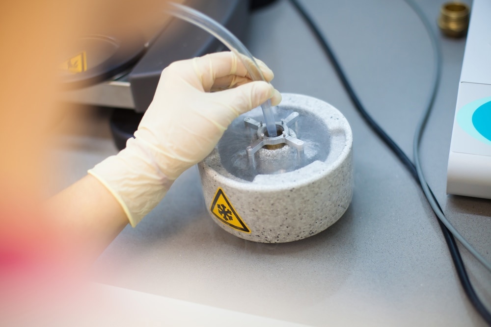
The process begins with vitrification; the biomolecule – usually a protein in solution – applied to a grid-mesh and plunge-frozen in liquid ethane. The sample is cooled so rapidly that the water molecules surrounding it don’t have time to crystallise, leading to an amorphous solid which has received little to no damage to the sample structure during the process. The sample is screened for particle concentration, distribution and orientation. A series of images are obtained and 2D classes are computationally extracted. The data is processed by reconstruction software which yields an accurate 3D model of the intricate biological structures at sub-cellular and molecular scales.
The technique, which was forty years in the making, won three scientists the Nobel Prize in Chemistry in 2017; Jacques Dubochet from the University of Lausanne in Switzerland, Joachim Frank from Columbia University in New York, USA, and Richard Henderson from the Medical Research Council Laboratory of Molecular Biology in Cambridge, UK. The technique can provide information on interactions otherwise impossible to visualise previously.
The greatest advantage of cryo-electron microscopy (cryo-EM) is its ability to analyse large, complex and flexible structures and is an alternative to X-ray crystallography of nuclear magnetic resonance (NMR) spectroscopy. This includes many biologically important proteins with variable or flexible structures, such as membrane proteins.
While X-ray crystallography can be used to obtain high-resolution structures, it requires that the molecules be crystallised; many proteins don’t fare well during the process as their structure is altered and therefore do not accurately represent the molecule, if they crystallise at all. Cryo-EM does not require crystals and allows scientists to observe biomolecules moving and functioning, which is much harder to do with crystallography.
NMR can also reveal very detailed structures but is restricted to relatively small proteins, or parts of them, including soluble, intracellular proteins rather than those embedded in cell membranes. Cryo-EM allows scientists to study larger proteins, including membrane-bound receptors or complexes of several biomolecules together.
Although its initial inception was delayed until advancements had been made in microscope design, imaging hardware, enhanced image processing and automation, cryo-EM has transformed structural biology. It has become one of the most critical tools in the laboratory for allowing scientists to determine the structure of biomolecules in solution at high-resolution.
References and further reading
Disclaimer: The views expressed here are those of the author expressed in their private capacity and do not necessarily represent the views of AZoM.com Limited T/A AZoNetwork the owner and operator of this website. This disclaimer forms part of the Terms and conditions of use of this website.