Aug 28 2008
An ultrasound probe small enough to ride along at the tip of a catheter can provide physicians with clearer real-time images of soft tissue without the risks associated with conventional x-ray catheter guidance.
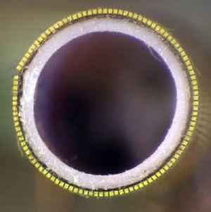
 3-D Transducer. Credit: Ned Light, Duke University
Duke University biomedical engineers designed and fabricated the novel ultrasound probe which is powerful enough to provide detailed, 3-D images. The new device works like an insect's compound eye, blending images from 108 miniature transducers working together.
Catheter-based procedures involve snaking instruments through blood vessels to perform various tasks, such as clearing arteries or placing stents, usually with the guidance of x-ray images.
In a series of proof-of-principle experiments in a water tank using simulated vessels, the engineers used the new ultrasound probe to guide two specific procedures: the placement of a filter within a vessel and the placement of a synthetic "patch" for aortic aneurysms. The scientists plan to begin tests of the new system in animals within the year.
"There are no technological barriers left to be overcome," said Stephen W. Smith, director of the Duke University Ultrasound Transducer Group and senior member of the research team that published the results of its latest experiments online in the journal IEEE Transactions on Ultrasonics, Ferroelectrics and Frequency Control. It is the cover article for the September issue.
"While we have shown that the new probe can work for two types of procedures, we believe that results will be more far-reaching," Smith said. "There are many catheter-based interventional procedures where 3-D ultrasound guidance could be used, including heart valve replacements and placement of coils in the brain to prevent stroke. Wherever a catheter can go, the probe can go."
Currently, when maneuvering a catheter through a vessel, physicians rely on x-ray images taken from outside the body and displayed on a monitor to manipulate their instruments. Often, a contrast agent is injected into the bloodstream to highlight the vessel.
"While the images obtained this way are good, some patients experience adverse reactions to the contrast agent," said research engineer Edward Light, first author of the paper and designer of the new probe. "Also, the images gained this way are fleeting. The 3-D ultrasound guidance does not use x-ray radiation or contrast agents, and the images are real-time and continuous."
Another benefit is portability, which is an important issue for patients who are too sick to be transported, since x-rays need to be taken in specially equipped rooms, Light said. The 3-D ultrasound machine is on wheels and can be moved easily to a patient's room.
Advances in ultrasound technology have made these latest experiments possible, the researchers said, by generating detailed, 3-D moving images in real-time. The Duke laboratory has a long track record of modifying traditional 2-D ultrasound – like that used to image babies in utero – into the more advanced 3-D scans. After inventing the technique in 1991, the team also has shown its utility in developing specialized catheters and endoscopes for real-time imaging of blood vessels in the heart and brain.
After testing many iterations of the design of the probe, also known as a transducer, the engineers came up with a novel approach – lining the front rim of the catheter sheath with 108 miniature transducers.
"These tiny transducers work together to create one large transducer, working much like the compound eyes of insects," Light explained.
In the first experiment, the new probe successfully guided the placement of a filter in a simulated vena cava, the large vein that carries deoxygenated blood from the lower extremities to the back to heart. Patients with clots in their legs – known as deep vein thrombosis – who cannot get clot-busting drugs often receive these filters to prevent the clots dislodging and traveling to the heart and lungs.
The second experiment involved the placement of abdominal aorta aneurysm stent grafts, which are large synthetic "tubes" used to patch weakened areas of the aorta that are at risk of bursting.
"I believe we have shown that 3-D ultrasound clearly works in a wide variety of interventional procedures," Smith said. "We envision a time in the not-too-distant future when this technology becomes standard equipment in various catheter kits."