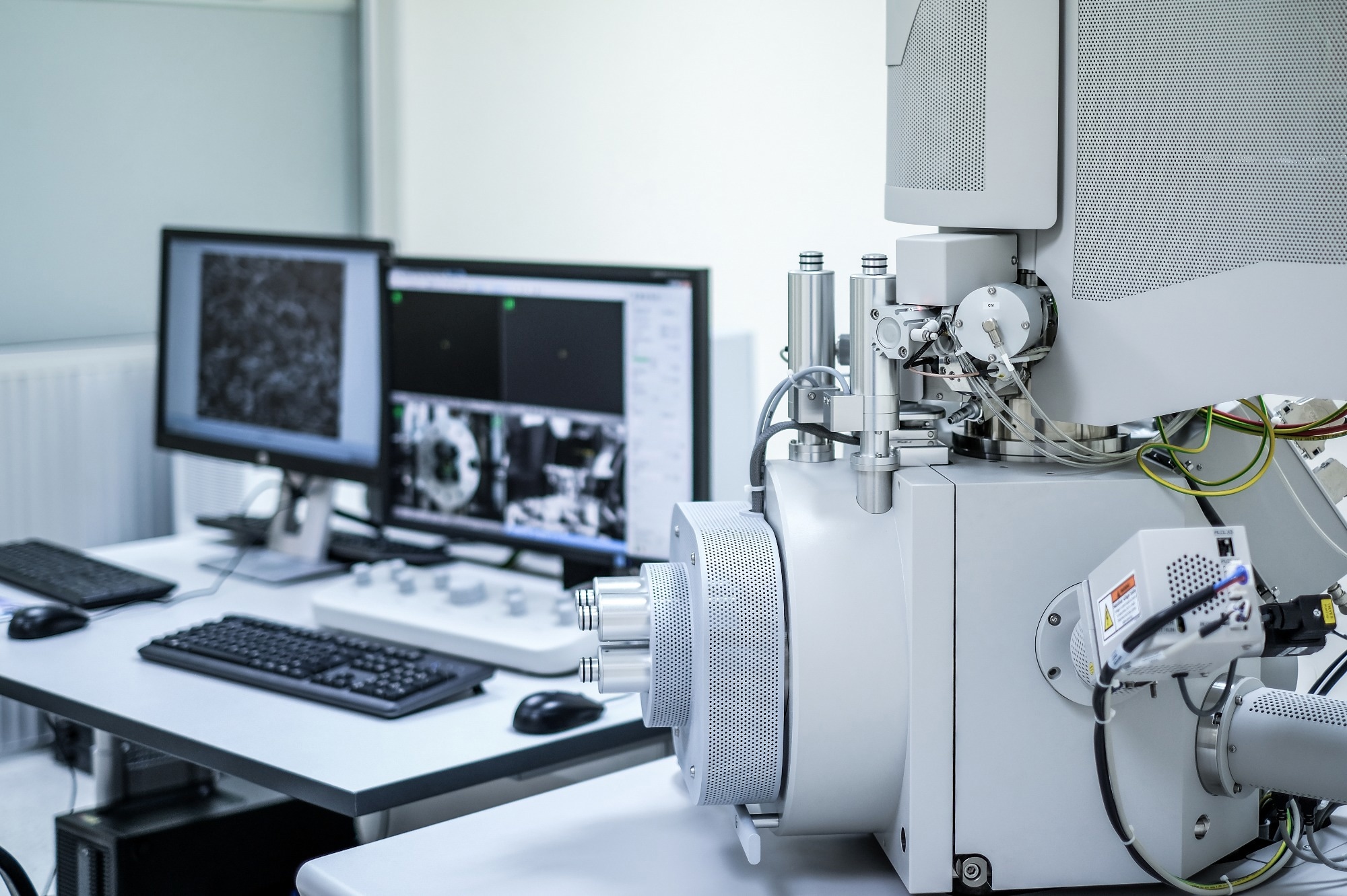
In a recent article published in Communications Engineering, researchers have introduced a novel method for automatically optimizing beam settings in scanning electron microscopy (SEM), significantly enhancing the accuracy and efficiency of nanoscale material analysis.
They developed an algorithm that eliminates the need for manual hardware adjustments, a frequent challenge in SEM operation, particularly in high-volume industrial environments.
 Study: Automatic beam optimization method for scanning electron microscopy based on electron beam Kernel estimation. Image Credit: Anucha Cheechang/Shutterstock.com
Background
SEM is crucial in materials science, nanotechnology, and semiconductor manufacturing. It uses a focused beam of electrons to scan a sample's surface, producing high-resolution images of its topography and composition.
The quality of these images depends on precisely controlling beam parameters like focus, stigmation, and aperture alignment. Traditionally, optimizing these parameters required skilled operators, making it a manual and time-consuming process.
In the semiconductor industry, SEM is extensively used for critical dimension measurements and defect inspection during device manufacturing. As semiconductor devices continue to shrink, there is an increasing demand for more precise and efficient SEM imaging techniques.
Therefore, automation in SEM operations is crucial in high-volume manufacturing environments, where throughput and consistency are critical.
About the Research
In this paper, the authors address the challenges of manual beam optimization in SEM by introducing a novel method for automatic beam parameter adjustment. Their approach uses beam kernels, a mathematical representation of the shape of the electron beam, to find the best settings for various SEM parameters.
They developed a computational model to simulate SEM image formation, considering the interaction between the electron beam and the sample surface.
Advanced image analysis techniques, including blind deconvolution, were employed to estimate the unknown surface image and the beam kernel from SEM images. This estimation process involves iteratively refining the beam kernel to minimize discrepancies between observed and simulated images.
The refined beam kernel is then used to determine correction values for key beam parameters such as focus, stigmata (x, y), and aperture alignment (x, y).
These correction values guide the automatic adjustment of the SEM's hardware components, eliminating the need for manual intervention.
To validate their approach, the researchers integrated the algorithm into an in-house software program capable of real-time SEM control. This software interfaces seamlessly with the SEM’s application programming interface (API), allowing for automated control over beam parameters and image acquisition.
Research Findings
The authors conducted experiments using various samples, including a reference gold on carbon (GoC) and two logic device semiconductor samples.
They demonstrated that their method effectively optimized beam parameters, achieving convergence to near-ground truth values within 15 iterations.
The average distances from the converged points to the ground truth values were 0.29 μm and 0.28 μm for focus, 0.34% and 0.08% for the stigmator and 0.17% and 0.19% for aperture alignment.
The SEM model used in the experiments featured a Gemini 2 column and double condenser lenses, maintaining high resolution even at high beam currents. Additionally, the researchers developed a custom C#-based auto-recipe program to interface with the SEM's API, enabling automated control over beam parameters and image acquisition.
Their method outperformed conventional sharpness-based methods in terms of accuracy and efficiency.
Applications
The developed automatic beam optimization method has significant implications for various domains. It is particularly beneficial in analytical laboratories where samples frequently change, requiring constant conditions adjustments.
Similarly, in focused ion beam (FIB)-SEM systems, where state variations necessitate regular readjustments after each milling step, this method could be invaluable.
While the focus of the research is on the semiconductor field, it can be applied in other areas, like biology, where it could reduce the time required for specimen preparation and observation.
Conclusion
In summary, the novel approach proved effective for automatically optimizing beams in SEM. Its ability to optimize multiple parameters simultaneously made it versatile and adaptable for various applications.
Additionally, it significantly enhanced the consistency of SEM analyses, particularly in industrial settings with high volumes of samples.
The authors acknowledged limitations and challenges and suggested that future work could focus on improving the method's accuracy and efficiency, especially at very high magnifications.
Furthermore, integrating this method with other advanced SEM techniques, such as electron backscatter diffraction (EBSD) and energy-dispersive X-ray spectroscopy (EDS), could lead to even more powerful analytical capabilities.
Journal Reference
Disclaimer: The views expressed here are those of the author expressed in their private capacity and do not necessarily represent the views of AZoM.com Limited T/A AZoNetwork the owner and operator of this website. This disclaimer forms part of the Terms and conditions of use of this website.