A digital holographic microscope's spatial resolution can be enhanced by incorporating structured illumination architecture. A study published in Optics and Lasers in Engineering employed this technique in a highly stable and cost-effective Fresnel biprism-based digital holographic microscope. The proposed setup enables high-resolution, three-dimensional imaging of living cells, and improves the spatial resolution by two folds.

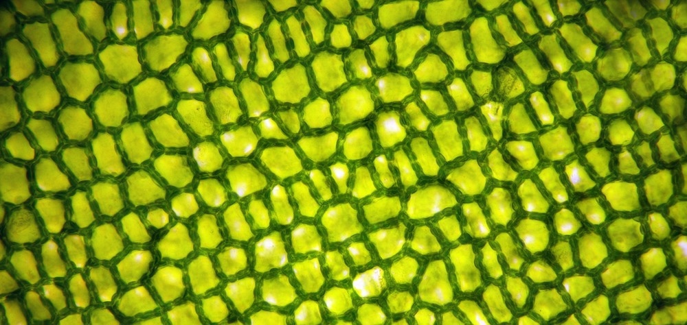
Study: Structured illumination in Fresnel biprism-based digital holographic microscopy. Image Credit: Barbol/Shutterstock.com
What is Digital Holographic Microscopy?
Holographic microscopy is a common quantitative tool for detailed cell analysis. With the development of high-resolution image sensors, holograms can be recorded digitally, allowing for non-invasive observation and analysis of living cells without damaging cell structure.
Digital holographic (DH) microscopy is an effective label-free biomedical imaging technique that provides complete access to the sample's amplitude and phase distribution.
The biochemical, mechanical, and morphological properties of microstructures and living cells can be studied simultaneously by developing desired DH conditions and integrating them with various imaging systems.
The Diffraction-Limited Lateral Resolution of DH Microscopy
Plane-wave illumination is often used in DH microscopy; the lateral resolution of the imaging setup is limited by its numerical aperture and wavelength. Shorter wavelength sources (X-rays or UV) can increase the lateral resolution. However, they can damage certain samples, including biological tissues.
A microscope with a large numerical aperture enables a larger numerical aperture. However, this results in a limited focus depth and working distance.
Structured illumination (SI) is a promising technique to enhance the imaging system's resolution by illuminating the sample in a periodic pattern. However, this approach is time-consuming, and mechanical vibrations can increase the temporal noise level.
SI implementation would benefit from the common-path geometry, as it ensures the stability of parameters over a long time.
Using Structured Illumination in Digital Holographic Microscopy to Enhance the Spatial Resolution
The traditional DH microscopy system is integrated with a SI approach employing a double-lens and grating system. The proposed methodology is a modified common-path configuration based on a biprism interferometer.
The modified configuration permits the use of structured light produced by a grating to improve the resolution of DH technologies. It involves two off-axis acquisitions of conventional illumination (CI) and synthetic illumination (SI) holograms.
The structured light brings the high-frequency components into the visible spectrum. As a result, a SI hologram has mixed low-frequency and high-frequency information. The conventional illumination hologram's spectrum is removed to ensure that the high-frequency components and the low-frequency information of the sample in the SI hologram spectrum do not overlap.
The hologram's amplitude is calculated using a numerical autofocusing technique and the angular spectrum propagation (ASP) method. After capturing two images, one for the CI pattern and the other for the SI pattern, the angular spectra of the holograms are digitally filtered and transmitted to create a high-resolution image of the sample.
Important Findings of the Study
A low-cost and highly stable method for imaging amplitude and phase distribution with a twofold enhancement in spatial resolution is developed by combining the SI pattern provided by a compact disk and the common-path off-axis geometry established by Fresnel biprism.
The experimental findings for a standard test sample verify the projected resolution enhancement over the traditional imaging system, as the proposed method successfully detects the cell's biconcave structure.
The lateral resolution prediction was tested using a standard test sample. The findings indicate that the SI system of biprism-based DH microscopy outperforms the CI-DH system by a quality factor of two.
The integrated SI and biprism interferometer setup needs two holograms without moving the grating, and its common-path design offers a very stable system for multi-shot imaging techniques.
Due to the close optical path length and the same intensity of the waves deflected by the biprism, high contrast and linear fringes are formed, enhancing the accuracy of measurements.
The proposed approach ignores the quadratic phase factor's impact on the rebuilt phase maps, leaving both beams with the same curvature. Therefore, the Fresnel biprism-based common-path configuration is an effective technique to eliminate undesired and uncorrelated phase fluctuations.
Furthermore, this quantitative DH system is suited for high-resolution 3D phase imaging because of its adjustable fringe modulation, ease of installation, and inexpensive cost.
The proposed DH technique provides high-resolution, more detailed imaging of live cells than standard DH microscopes. In addition, it can be linked with various optical microscopes to produce label-free multimodal imaging.
Reference
Yaghoubi, S. H. S., Ebrahimi, S., & Dashtdar, M. (2022). Structured illumination in Fresnel biprism-based digital holographic microscopy. Optics and Lasers in Engineering. https://www.sciencedirect.com/science/article/abs/pii/S0143816622002688
Disclaimer: The views expressed here are those of the author expressed in their private capacity and do not necessarily represent the views of AZoM.com Limited T/A AZoNetwork the owner and operator of this website. This disclaimer forms part of the Terms and conditions of use of this website.