Feb 18 2020
When one looks via a microscope, any object located on the stage is enlarged to such an extent that it can hardly be imagined by the naked eye.
 UConn researchers plan to continue to refine the technology to enhance its use in commercial and clinical applications. Image Credit: Guoan Zheng.
While conventional microscopy techniques bring tiny details into view, regular instruments do not offer a complete picture.
A majority of the optical microscopes have a restricted field of view, which is just 1 to 2 mm. This poses a major difficulty to pathologists and life scientists who depend on microscopy to diagnose and analyze various diseases because prepared samples of tissues have sized down to the centimeter range.
The University of Connecticut (UConn) has now developed a novel microscopy platform to deal with this unmet clinical need. But this microscopy platform does not include objective lenses, the core component of conventional microscopes. With a lensless platform, scientists can truly offer a more complete picture to clinicians and thus guide them to make more precise diagnoses.
Guoan Zheng, a professor of biomedical engineering at the University of Connecticut, has recently reported his study findings in the Lab on a Chip journal, following an effective demonstration of a lensless on-chip microscopy platform.
This unique platform removes several of the most standard issues associated with traditional optical microscopy and offers a cost-effective option for diagnosing the disease.
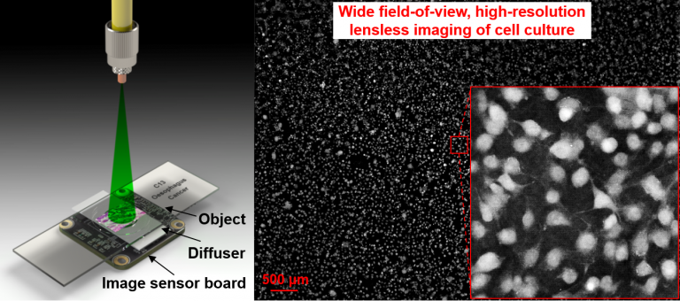
Instead of utilizing lenses to enlarge the tissue sample, the new platform developed by Zheng depends on a diffuser that goes between the camera/image sensor and the specimen. This diffuser arbitrarily moves to varying positions, and the sensor simultaneously obtains the images, collecting information on the encoded object that will be subsequently utilized to recover an image for viewing by researchers or clinicians.
An imaging technique known as ptychography is the core of the object recovery process. Generally, ptychographic imaging utilizes a focused beam to light up a sample and captures the pattern produced by the diffracted light.
To recover a complete complex image, such as a tissue sample, for viewing purposes, ptychography involves recording an unlimited number of patterns while scanning the sample to different positions.
Although ptychography has been of increasing interest to scientists around the world, broad implementation of the method has been hampered by its slow speed and the requirement of precise mechanical scanning.
Shaowei Jiang, Study Lead Author and Graduate Student, University of Connecticut
The new ptychographic technology developed by Zheng tackles these problems by bringing the sample nearer to the image sensor. The latest configuration enables the researchers to set the whole area of the image sensor as the imaging field of view. Moreover, it eliminates the precise mechanical scanning required for standard ptychography.
The reason is the latest configuration has the Fresnel number of around 50,000, which is the highest to be ever tested for ptychography. The Fresnel number defines the way a light wave moves across a distance after traveling via an opening, such as a pinhole.
Zheng’s experiments made use of the ultra-high Fresnel number, which indicated that there were very minimal diffraction of light from the object plane to the sensor plane. Furthermore, reduced levels of diffraction implied that the diffuser’s motion could be directly monitored from the captured raw images. This removed the need for an accurate motion stage, which is crucial for standard ptychography.
This approach cuts down on processing time, cost, and allows for a more complete image to be produced of the sample.
Guoan Zheng, Professor, Biomedical Engineering Department, University of Connecticut
With traditional lensed microscopy, researchers can only visualize a tiny part of a slide during each viewing process. The platform developed by Zheng provides a considerable enhancement by effectively widening the field of view of the microscope.
The present prototype of Zheng provides a field of view of 30 mm2, as opposed to the regular ~2 mm2. By utilizing a full-frame image sensor in a standard photography camera, Zheng’s technology enables physicians to examine two entire slides simultaneously.
“Imagine being able to read a whole book at once instead of just a page at a time. That’s essentially what we hope our technology will allow clinicians to do,” Zheng added.
Zheng’s new platform adds to its already long list of enhancements and removes the need to stain cells. Researchers would usually stain portions of cells, just like the nucleus, to detect the number of cells present. Zheng tested the potential of this platform to execute automatic cell segmentation utilizing the recovered label-free phase maps.
Owing to the rugged performance and compact configuration of the platform, Zheng and his group believe that their prototype would be suitable for a wide range of telemedicine, global health, and point-of-care applications. The novel technology can even be useful for electron and X-ray microscopy.
By using our lensless, turnkey imaging system, we can bypass the physical limitations of optics and acquire high-resolution quantitative information for on-chip microscopy. We’re excited to continue to refine this technology for commercial and clinical applications to have a tangible impact for patients and researchers.
Guoan Zheng, Professor, Biomedical Engineering Department, University of Connecticut
“Wide-field, high-resolution lensless on-chip microscopy via near-field blind ptychographic modulation” is the title of the study published in the Lab on a Chip journal. The study was financed by the National Science Foundation, grant #1510077.