Aug 19 2019
Scientists have created an innovative self-calibrating endoscope that generates 3D images of objects that are smaller than a single cell.
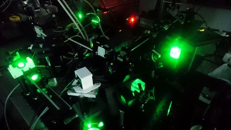
 Researchers have developed a new self-calibrating endoscope that produces 3D images of objects smaller than a single cell. (Image credit: J. Czarske, TU Dresden, Germany)
The tip of the endoscope—with no lens or any optical, mechanical, or electrical parts—measures just 200 μm across, about the thickness of a few human hairs twisted together.
Being a minimally invasive tool for imaging inner features of living tissues, the very thin endoscope could enable a range of research and medical applications. The study will be presented at the Frontiers in Optics + Laser Science (FIO + LS) conference, conducted from September 15th to 19th, 2019, in Washington, D.C., the United States.
The lensless fiber endoscope is approximately the size of a needle, allowing it to have minimally invasive access and high-contrast imaging as well as stimulation with a robust calibration against bending or twisting of the fiber.
Juergen W. Czarske, Study Lead Author, Director and C4-Professor, TU Dresden, Germany
The endoscope will possibly be valuable specifically for optogenetics—research strategies that use light to trigger cellular activity. Additionally, it could prove valuable for monitoring cells and tissues during medical procedures and also for technical inspections.
A Self-Calibrating System
Traditional endoscopes use cameras and lights to take images within the body. Recently, scientists have created alternative methods to take images via optical fibers, eliminating the need for bulky cameras and other bulky parts, resulting in considerably thinner endoscopes. However, despite their potential, these technologies experience restrictions such as the incapability to withstand temperature variations or bending and twisting of the fiber.
One main obstacle to making these technologies practical is that they need intricate calibration processes, in several cases when the fiber is collecting images. To tackle this, the scientists incorporated a thin glass plate, with a thickness of just 150 μm, to the tip of a coherent fiber bundle, a type of optical fiber that is generally used in endoscopy applications. The coherent fiber bundle used in the experiment had a width of 350 μm and included 10,000 cores.
Upon illuminating the central fiber core, it emits a beam that is reflected back into the fiber bundle and acts as a virtual guide star for quantifying how the light is being transmitted, called the optical transfer function. The optical transfer function offers vital data used by the system to calibrate itself on the go.
Keeping the View in Focus
A spatial light modulator is an important part of the new setup, which is used to control the direction of the light and allow remote focusing. The spatial light modulator compensates the optical transfer function and images onto the fiber bundle. The camera captures the back-reflected light from the fiber bundle and is superposed with a reference wave to measure the light’s phase.
The position of the virtual guide star reveals the instrument’s focus, with a minimal focus diameter of nearly 1 μm. The scientists employed an adaptive lens and a 2D galvanometer mirror to shift the focus and allow scanning at various depths.
Demonstrating 3D Imaging
The group tested their device by using it to image a 3D specimen beneath a 140-μm thick cover slip. The device scanned the image plane in 13 steps over 400 μm with an image rate of 4 cycles per second and successfully imaged particles at the top and bottom of the 3D specimen.
However, its focus weakened with the increase in the angle of galvanometer mirror. The scientists suggest that further research could overcome this challenge. Moreover, using a galvanometer scanner with a higher frame rate could enable more rapid image acquisition.
The novel approach enables both real-time calibration and imaging with minimal invasiveness, important for in-situ 3D imaging, lab-on-a-chip-based mechanical cell manipulation, deep tissue in vivo optogenetics, and key-hole technical inspections.
Juergen W. Czarske, Study Lead Author, Director and C4-Professor, TU Dresden, Germany