Aug 5 2019
Scientists describe a new imaging system that cancels chromatic optical aberrations present in the eye of certain people, thereby enabling a more accurate evaluation of vision and eye health.
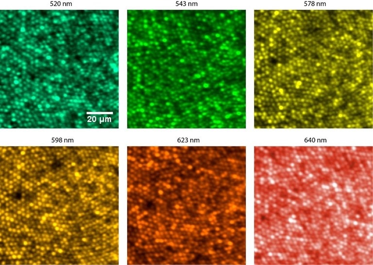
 Images of the smallest cone photoreceptors in the retina, about 2 µm wide. Coloration was added to represent the different wavelengths of light used to capture the images after compensating chromatic aberration. (Image credit: Xiaoyun Jiang and Ramkumar Sabesan, University of Washington)
By clicking pictures of the eye’s tiniest light-sensing cells with various wavelengths, the system also offers the first objective measurement of longitudinal chromatic aberrations (LCA), which could result in a new understanding of their relationship to glare visual halos, and color perception.
In Optica, The Optical Society’s journal for high-impact research, the scientists, from the University of Washington, Seattle, U.S.A., say the technology can be freely deployed in the clinic, where it could be most useful for evaluating changes in the eye associated with aging and can also help inform the design of new multifocal lenses by taking into consideration chromatic aberrations in the lenses themselves. For vision research, the method could progress studies on color blindness and how different people distinguish color.
The previous methods of compensating the eye’s native LCA rely on population average estimates, without individualized correction on a person-by-person basis. We demonstrate a modified filter-based Badal optometer that offers the capability to tune LCA across different wavelength bands and for each individual in a customized fashion.
Ramkumar Sabesan, Study Team Leader, University of Washington, Seattle
The scientists describe adding a new optical assembly into conventional adaptive optics instruments to create individually customized high-resolution, multi-wavelength pictures of the smallest cone photoreceptors in the eye, measuring about 2 µm across.
Our study establishes a flexible tool to compensate for chromatic aberration in different wavelength bands and in an individualized manner, thus facilitating future investigations into how we see color in our environment, unimpeded by the native chromatic imperfections of the individual. Now equipped with the tools to control chromatic aberration, we plan to conduct studies on normal and deficient color vision.
Ramkumar Sabesan, Study Team Leader, University of Washington, Seattle
Compensating for aberrations
Similar to man-made optical elements such as camera lenses and microscopes, the cornea and lens of the eyeball have optical aberrations that blur the image formed on the retina. Aberrations distort the images projected on a person’s retina, diminishing his/her vision. They also influence the images doctors gain when viewing the inside of the eye using ophthalmologic tools.
Adaptive optics is a technique to make up for these aberrations. Adaptive optics technology, presently used by astronomers to resolve aberrations that happen when viewing space through Earth’s atmosphere, have been integrated into eye imaging instruments. However, while present-day instruments are successful at rectifying monochromatic aberrations (those that do not vary based on the wavelength of light being applied), chromatic aberrations (those that are influenced by wavelength) are more difficult.
To overcome this issue, present-day instruments use assumptions about the aberrations anticipated in an average or “typical” eye, instead of information about the actual aberrations in a particular person’s eye. While this is adequate for a number of applications, it is less appropriate for other applications that demand the simultaneous and fine focus control of various wavelengths.
To resolve this drawback, the scientists used a device called a Badal optometer, which comprises a pair of lenses that are a specific distance apart. Altering the distance between the two lenses alters the focus without changing the size of an image viewed via the lenses.
The scientists altered this basic Badal optometer by incorporating two filters that convey longer wavelengths of light while reflecting shorter ones. These filters were kept immobile within a traditional Badal optometer, such that now, when the distance between the lenses is altered, the conveyed and reflected wavelength bands have subtly varying levels of focus enough to compensate for the eye’s native chromatic aberration for the two wavelength bands.
By excellently tweaking the selection of filters, distances between the lenses, and multi-color illumination, this configuration can be used collectively to measure and compensate for chromatic aberration in a tailored manner.
A valuable tool for the clinic and the laboratory
The scientists set up their new LCA compensator in two different adaptive optics instruments: adaptive optics vision simulation and adaptive optics scanning laser ophthalmoscopes. The eyes of human volunteers were imaged through these new instruments.
They learned that the new technique effectively overcame discrepancies in earlier estimates of the human eye’s native LCA related to monochromatic aberration, depth of focus, and wavelength-reliant light interactions with retinal tissue. When monochromatic as well as chromatic aberrations were compensated, a person’s vision was limited only by the arrangement of cone photoreceptors—light-detecting cells—in the retina, while eliminating the chromatic aberration compensation meant that either green or red vision was enhanced.
The scientists also showed the system’s capacity to image the tiniest cone photoreceptors with several wavelengths concurrently by reducing chromatic aberration, establishing that the Badal LCA compensator provides a fine level of detail, a crucial advancement for supporting color vision research.
Besides providing improved images of the inside of the retina, the technology is beneficial for examining how chromatic aberrations impact visual performance and retinal image quality. This has earlier been tough because instruments that offer fine individualized control of LCA were not available. Furthermore, measurements of LCA acquired subjectively and objectively did not meet the expectations.
By applying the technology to two different adaptive optics-based modalities, we show a high fidelity of visual performance and retinal imaging once chromatic and monochromatic aberrations are compensated. The high-resolution retinal images thus obtained allowed us to quantify chromatic aberration objectively and compare against a large body of literature dedicated to measuring chromatic aberration.
Ramkumar Sabesan, Study Team Leader, University of Washington, Seattle
Scientists stated that the new system is all set for deployment in adaptive optics instruments that are already regularly used in the clinic and for translational research.