Jul 1 2019
A portable and low-cost optical coherence tomography, or OCT, scanner has been developed by biomedical engineers at Duke University. The tool has great potential to bring vision-saving technology to underserved areas, both across the United States and abroad.
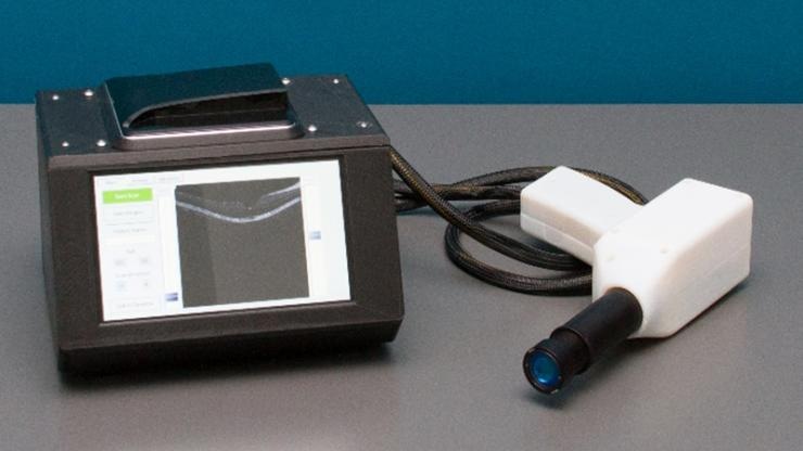
 This OCT system designed at Duke University is 15 times lighter and smaller than current commercial systems and is made from parts costing less than a tenth the retail price of commercial systems. (Image credit: Duke University)
The innovative scanner is 15 times smaller and lighter than present-day commercially available systems and this can be attributed to a remolded, 3D-printed spectrometer. It is fabricated from parts that cost less than one-tenth the retail price of systems available in the market and yet does not affect the imaging quality.
The novel OCT scanner, in its initial clinical trial, generated images of 120 retinas that were 95% as sharp as those captured by existing commercially available systems. Such vivid sharpness was enough for precise clinical diagnosis.
The study results have been published online in Translational Vision Science & Technology, an ARVO journal, on June 28th, 2019.
OCT imaging has been in use since the 1990s, and over these years, it has become the standard of care for diagnosing a number of retinal diseases such as diabetic retinopathy and macular degeneration, and also for glaucoma. Conversely, OCT is seldom included as part of a standard screening exam because these machines are exorbitantly priced, costing over $100,000—implying that most often they are available in only larger eye centers.
Once you have lost vision, it’s very difficult to get it back, so the key to preventing blindness is early detection. Our goal is to make OCT drastically less expensive so more clinics can afford the devices, especially in global health settings.
Adam Wax, Professor, Biomedical Engineering, Duke University
The optical analog of ultrasound OCT functions by relaying sound waves inside tissues and determining the duration taken by them to come back. However, since light is relatively faster than sound, it is not easy to measure time. In order to time the light waves reflecting back from the tissue being scanned, OCT devices employ a spectrometer to establish how much their phase has moved as opposed to identical light waves, which although have not interacted with tissue, have traveled the same amount of distance.
The main technology allowing the smaller and less costly OCT device is a new kind of spectrometer created by Wax and Sanghoon Kim, his former graduate student. Conventional spectrometers are mostly made of accurately cut metal components and direct light via an array of mirrors, lenses, and diffraction slits that are shaped like the alphabet W. Although this setup offers a high degree of precision, mild mechanical shifts induced by bumps or even temperature variations can result in misalignments.
However, Wax’s design takes the light on a spherical path inside a housing which is largely made from 3D-printed plastic. Since the light path of the spectrometer is circular, any contractions or expansions owing to temperature variations take place symmetrically, thus balancing themselves out so that the optical elements remain aligned. In addition, the device employs a larger detector by the end of the light’s journey to eliminate misalignments as much as possible.
Conventional machines weigh over 60 pounds and occupy an entire desk. They also cost anywhere between $50,000 and $120,000. By contrast, the latest OCT device weighs only four pounds, is roughly the size of a lunch box, and will be sold for below $15,000, Wax believes.
Right now OCT devices sit in their own room and require a PhD scientist to tweak them to get everything working just right. Ours can just sit on a shelf in the office and be taken down, used and put back without problems. We’ve scanned people in a Starbucks with it.
Adam Wax, Professor, Biomedical Engineering, Duke University
In the latest research, J. Niklas Ulrich, retina surgeon and associate professor of ophthalmology at the University of North Carolina School of Medicine, tested the novel OCT scanner against a commercially available instrument developed by Heidelberg Engineering. To do this, he carried out clinical imaging on both eyes of 60 patients—half from volunteers with some kind of retinal disease and half from healthy volunteers.
Then to compare the images created by both machines, the team utilized contrast-to-noise ratio—a measure that is largely used for determining the quality of images in medical imaging. The outcomes showed that, through this metric, the portable and low-cost OCT scanner provided valuable contrast that was just 5.6% less than that of the commercially available machine, which is still sufficiently good to enable clinical diagnostics.
I have been very impressed by the quality of images from the low-cost device—it is absolutely comparable to our standard commercial machines. It obviously lacks some bells and whistles of our $100k+ OCT scanners, but allows for accurate diagnosis of structural retinal disease as well as monitoring of treatment success. The setup is quick and easy with a small footprint, allowing the device to perform well in smaller satellite offices. I hope that the development of this low-cost OCT will improve patients’ access to OCT technology and contribute to saving sight in North Carolina as well as nationally and worldwide.
J. Niklas Ulrich, Retina Surgeon and Associate Professor, Ophthalmology, University of North Carolina School of Medicine
At present, Wax is commercializing the device via a startup company known as Lumedica, which is already creating and marketing first-generation instruments for use in research. The company is aiming to secure venture backing in the coming days and is also in discussion to obtain prospective licensing deals with outside firms.
“There’s a lot of interest from people who want to take OCT to new parts of the globe as well as to underserved populations right here in the U.S.,” stated Wax. “With the growing number of cases of diabetic retinopathy in places like the United States, India and China, we hope we can save a lot of people’s sight by drastically increasing access to this technology.”
The National Institutes of Health (R01 CA210544) and the Duke-Coulter Translational Partnership funded the study.
Low-Cost OCT System
(Video credit: - Duke University)