Oct 28 2015
Researchers from the Paul Scherrer Institute PSI have succeeded in using commercially available camera technology to visualise terahertz light. In doing so, they are enabling a low-cost alternative to the procedure available to date, whilst simultaneously increasing the comparative image resolution by a factor of 25.
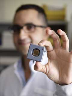
 Mostafa Shalaby with the CCD sensor. Its resolution is 25 times higher than that of the sensors used to visualise terahertz light up to now. (Photo: Paul Scherrer Institute/Markus Fischer)
The special properties of terahertz light make it potentially advantageous for many applications, from safety technology to medical diagnostics. It is also an important tool for research. At PSI, it will be used for the experiments on the X-ray free-electron laser SwissFEL. The terahertz laser developed at PSI is currently the world’s most intensive source of terahertz light. The researchers are presenting their findings in the scientific journal “Nature Communications”.
From security controls to the detection of tumours: If you want to uncover hidden structures, terahertz light is a method with potential. It penetrates paper, plastic or textiles easily and renders any objects hiding behind them visible. And although its penetration depth in biological tissue is only a few millimetres, it has one property that also makes it particularly interesting for medical diagnostics: Unlike the radiation from an X-ray, it leaves the tissue unharmed.
The reason it’s so harmless is that terahertz light is composed of particles (photons) that are relatively low in energy. This property is advantageous for research, where it helps to trigger processes without the trigger itself leaving behind any trace. One example is research into new materials for magnetic data storage, where flashes of terahertz light can be used to instantaneously change the magnetisation or the optical properties of the material to be examined.
Too gentle for strong sensors
As practical as this property of terahertz light is, it is specifically its low photon energy that causes a few issues. Until recently, it was only possible to visualise terahertz light using what are referred to as bolometers. These sensors are not only expensive, they are also very sensitive to environmental influences – particularly to heat. Simply moving your hand too close to the sensor can be enough to falsify the outcome. In addition, the image resolution achievable is comparatively low.
Likewise, the CCD sensors already widely used in research and which are also incorporated in smartphones and video cameras for a razor sharp image could not be used for terahertz light to date. Since the terahertz light was too weak for the robust CCD sensors – the sensors simply did not respond to the light.
More intensity for a sharper image
Thanks to a stronger terahertz laser developed at PSI, Christoph Hauri and his team of researchers at PSI have now succeeded in overcoming the sensitivity threshold of the CCD sensors. “Unlike the terahertz laser sources available in the past, the terahertz laser developed at PSI is characterised by its particularly high intensity”, explains Christoph Hauri.
The researchers used their experiment to demonstrate that the intensive terahertz light created at PSI using a commercially available CCD sensor can be made visible. This is a major technological milestone: “Now that the terahertz light is intensive enough to be able to visualise it with a normal CCD sensor, we are getting images with a resolution that is 25 times better than with the bolometer”, explains Mostafa Shalaby, who carried out the experiment, “because the pixel size of the CCD sensor is only around one fifth of that of the bolometer.” And now that the CCD sensor can be used, another significant advantage comes fully into play: Its sensitivity to environmental influences like heat is negligible.
No longer groping in the dark
The intensive PSI terahertz laser was developed specially for future applications on the SwissFEL. The X-ray free-electron laser, SwissFEL, is currently under construction at PSI and will be commissioned from the end of 2016. It will produce X-ray light impulses with laser-like properties. The visualisation of terahertz light that is now possible using CCD sensors will result in a range of advantages. The terahertz lasers will be used in combination with the X-ray light of the SwissFEL. For example, to carry out research into new materials for magnetic data storage, a terahertz laser pulse triggers a change in the magnetisation of a sample of the material to be examined. The X-ray laser pulse of the SwissFEL then visualises the sample just a few femtoseconds later. This makes it possible to determine what happened in the sample during those femtoseconds.
The researchers are especially interested in the fact that the CCD sensors can now visualise terahertz light in its experimental environment. “This allows us to record the precise spatial position of the terahertz beam during the experiment”, emphasises Hauri. In addition, the refresh rate of the CCD sensor is high enough to keep up with the speed at which the experiments on the SwissFEL take place. The SwissFEL fires 100 X-ray light pulses per second, and a separate experiment is carried out with each of those pulses.
Now that the researchers have shown that the visualisation of terahertz light with CCD sensors works in principle, their aim is to develop this idea further. “It is of course possible to tailor CCD sensors for certain applications in research”, says Carlo Vicario, who carried out the experiment together with Mostafa Shalaby. The researchers also see great potential for applications beyond the field of research. As Hauri says: “CCD sensors are low cost and robust.” But for widespread use it would of course also be necessary to have marketable terahertz lasers of sufficient intensity. “CCD sensors not only perform better but also cost less than one tenth of the cost of a bolometer, they are sure to quickly gain a foothold in the fast-growing terahertz sector of science”, Hauri is convinced.
Background: A light with potential and pitfalls
With a wavelength of between 0.1 and 1 millimetre, terahertz light occupies a range between infrared radiation and microwave radiation. Unlike X-ray radiation, terahertz light is non-ionising, which is why it is believed based on current knowledge to cause less cell damage. That is of major importance for medical applications in particular.
Previously, the generation of usable terahertz light was hampered by weak beam power. Also, it only worked at very low temperatures. The terahertz laser developed at PSI is currently the world’s most intensive source of terahertz light. It achieves its intensity through a salt crystal called DAST which is irradiated using a strong infrared laser. This results in a mix of different frequencies that creates the terahertz light. The method used at PSI converts ten times more light into terahertz light than other methods. That is why it is more intensive. Also, the laser operates at room temperature.
The bolometers used so far to visualise terahertz light are based on measuring the change of heat. This makes them particularly sensitive to ambient heat influences. In addition, the image resolution achievable of 320 x 240 pixels with a pixel size of 23.5 micrometres is comparatively low.
The CCD sensor (CCD stands for “charge-coupled device”) used for the experiment had an image resolution of 1360 x 1024 pixels. Its pixel size was 4.65 micrometres. As a result, the images are considerably sharper than those generated using a bolometer.