Oct 29 2013
If you've ever been sleep-deprived, you've probably had a firsthand glimpse of the blood vessels in your eyes. But what you haven't seen—and what many eye care professionals cannot see as well as they would like—are the vessels closest to the retina, the light-sensitive tissue at the back of the eye.
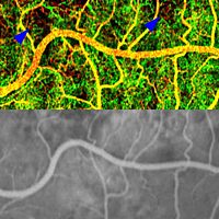
 Phase-variance OCT (top) can reveal blood vessels over the retina (yellow and red) and underneath it in a layer of tissue called the choriocapillaris (green). In this person with AMD, it also reveals areas of choriocapillaris loss (blue arrowheads). This level of detail isn't seen by standard fluorescein angiography (bottom). Republished from Kim et al., PNAS.
The National Eye Institute, part of the National Institutes of Health, is helping researchers develop new retinal imaging methods to solve this problem. Such methods have the potential to improve diagnosis and treatment of common eye diseases like age-related macular degeneration (AMD) and diabetic retinopathy. These diseases can cause a variety of defects in blood vessels inside the eye, including vessel loss, the growth of abnormal vessels, and small bulges that can rupture (called aneurysms).
"It makes sense to diagnose and monitor these conditions as closely as possible so that we can provide timely treatment," said Alfredo Dubra, Ph.D., an assistant professor of ophthalmology and biophysics at the Medical College of Wisconsin. "The main goal is to avoid irreversible damage," he said.
Since the 1960s, the gold standard test for viewing abnormal blood vessels in the eye has been fluorescein angiography. If an eye care professional detects signs of blood vessel abnormalities during a comprehensive dilated eye exam, this test is often the next step. Fluorescein is a dye that glows green under fluorescent light. In the test, it is typically injected into an arm vein. A camera is then used to take pictures as the dye moves through bloodstream into the eye, making it possible to see abnormal or damaged vessels there.
Clinicians and researchers are especially interested in seeing blood vessels nearest to the retina, because those vessels may show early signs of distress in some diseases. But conventional fluorescein angiography isn't powerful enough to see small retinal vessels and capillaries in fine detail.
From star gazing to eye gazing
Adaptive optics (AO) is one technology helping to overcome this problem. It deals with the tendency of light to become distorted as it passes through different media such as air, water, or living tissue. AO was originally developed because astronomers wanted to get a clear view of objects in space, without distortions caused by the atmosphere. But vision scientists and eye care experts have a similar problem: When light is shined into the eye, it is distorted by the cornea (the front, transparent part of the eye) and the lens. AO systems typically use a sensor to measure the pattern of blurring and a flexible mirror to correct it.
In theory, AO can be used to supercharge almost any type of retinal imaging. So Dubra collaborated with Richard Rosen, M.D., at the New York Eye and Ear Infirmary in order to build a device incorporating AO with fluorescein angiography. In a recent NEI-funded study involving 10 healthy volunteers, they found that the combined technology enabled them to see capillaries, tiny aneurysms, and other fine details of retinal blood vessels that aren't visible by standard fluorescein angiography.
Dubra and Rosen also tried another twist that could save scores of people from unpleasant reactions to fluorescein injections, which range from nausea to severe allergic reactions. In some sessions, the researchers gave volunteers an oral form of the dye mixed with orange juice. Although one person became nauseated after getting the typical dye injection, none of the subjects reported illness after drinking the dye.
The oral dye even gave an added boost to the test. "Drinking the dye turns out to provide a stronger signal that lasts longer," Dubra said.
From sound waves to light waves
Researchers are also working to enhance another method called optical coherence tomography (OCT). OCT itself has only been around since the early 1990s. It's similar to ultrasound, which creates images of living tissues by bouncing sound waves off of them. The difference is that OCT uses light waves instead. A painless infrared beam is scanned across the eye multiple times, creating virtual slices that are each less than one-tenth the thickness of a human hair. These slices can be taken at almost any orientation, making it possible to view the retina and other tissues in cross-section rather than face-on.
The retina viewed by phase variance-OCT.
John S. Werner, Ph.D., a professor of ophthalmology and neuroscience at the University of California, Davis, is working on combining AO with OCT. He's also developed a new version of OCT called phase-variance OCT.
"With phase-variance OCT, instead of acquiring each slice once, we acquire every slice three times in a row, and then we align them and ask what's different," Werner said. When there is motion between the three slices—for example, the flow of blood through veins and arteries—it creates contrast and enhances the composite image, he said.
In an NEI-funded study, Werner and colleagues used phase-variance OCT to examine the eyes of two volunteers in their 60s. One had no history of eye disease and the other had a type of advanced AMD called geographic atrophy. The researchers were able to see retinal vessels, as well as small vessels in a layer of tissue underneath the retina called the choriocapillaris.
In the person with AMD, they detected a loss of these small choroidal vessels, which typically aren't visible with fluorescein angiography or other standard methods.
Looking ahead
It may be a few years before phase-variance OCT and AO-enhanced imaging reaches your eye care professional's office. But experts say that these technologies will quickly expand into more research settings and into clinical trials, where they may enable more efficient testing of new treatments.
"For AMD and other eye diseases, these technologies have the potential to resolve small changes in the eye that occur as vision slowly deteriorates. That could help investigators monitor clinical trial participants more closely and detect the effects of new drugs much faster," said Matthew McMahon, Ph.D., senior adviser for translational research at NEI.
Werner and Dubra and their colleagues custom-built their imaging systems, but they're already thinking about how other researchers can adopt them. Standard OCT equipment can be modified to perform phase-variance OCT by adding new software, Werner said. The system from Dubra's group requires unique hardware, but they have a plan for gradually introducing it to the broader research community.
"We have a small network of collaborators that we've built instruments for and we're going to roll out the technique over the next year," Dubra said. These collaborators include Moorfields Eye Hospital in London; the University of Pennsylvania in Philadelphia; University of California, San Diego; and the NEI intramural research program in Bethesda, Md., he said.
The studies described here were supported in part by NEI grants EY017607, EY001931 and EY014743. NEI leads the federal government's research on the visual system and eye diseases. NEI supports basic and clinical science programs that result in the development of sight-saving treatments.