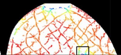
Tufts University School of Engineering has developed an innovative optical imaging technology that will assist doctors in identifying as well as monitoring a patient’s response to the primary treatment of breast cancer.
 Shown is an optical mammography image of hemoglobin oxygenation of a duct carcinoma in situ (DCIS), a breast cancer in the lining of the milk ducts that has not yet invaded nearby tissues. In this image, the boxed area corresponds to the cancer location and indicates lower values of hemoglobin oxygenation. Credit: Sergio Fantini, Ph.D., professor of biomedical engineering at Tufts University
Five years of clinical study at the Tufts and a grant of $3.5 million from the National Institutes of Health have gone into the development of this technology. The study is still under progress.
NIR (near infrared) light is used in this non-invasive technology to scan the breast tissue and an algorithm is subsequently applied to interpret the information. Variation in light absorption permits the identification of fats, water and oxygen-poor and oxygen-rich tissue, the main structures in a breast tissue.
Professor of Biomedical Engineering, Sergio Fantini stated that though x-ray mammography is good at finding out lesions, it gives compromised results when it comes to determining whether the suspicious lesions are cancer or not. The NIR technique of Tufts could complement typical mammography, mainly for women less than 40 years old, who have thick breast tissue which leads to obstruction of details in x-rays.
The application of the NIR technique for multiple usages within a short span of time is possible without the threat of radiation exposure as ionizing radiation is not used. One more advantage of this new technique is that it can virtually get immediate images of metabolic changes of hemoglobin concentration and oxygenation levels.
Optical mammography is more comfortable than conventional mammograms, as the breasts of the patient are lightly compressed amid two horizontal platforms made of glass. Real time images are displayed through the application of a specialized software program.