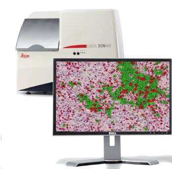
Leica Microsystems announces the release of Tissue IA 2.0, high performance image analysis for discovery research. Combining fluorescence and brightfield analysis capabilities in a single platform, with precision cell modelling, Tissue IA 2.0 offers a superior solution for IHC biomarker quantification.
Tissue IA 2.0 joins the Total Digital Pathology portfolio from Leica, providing streamlined end-to-end excellence in capture, management and analysis of digital pathology images.
 Tissue IA – Automated Image Analysis for Brightfield and Fluorescence Digital Pathology.
A major challenge in research today is retrieval of quantitative, reproducible data from tissue-based IHC studies. Tissue IA 2.0 provides expert tools for researchers to extract the most from their studies. Powerful color separation and multi-marker colocalization functionality provides advanced insight and unbiased measurement of multiple antigen immunostaining in brightfield or fluorescent samples. Sophisticated cell modelling accurately detects and quantifies differential expression of staining in cellular compartments, providing detailed insight into cytoplasmic, membrane and nuclear biomarker localization.
The advanced dual staining capabilities in Tissue IA 2.0 enable researchers to identify cell cohorts at the molecular level. Use one marker to identify a population of interest and then quantify expression of a second, providing exceptional analysis performance and greater understanding of a user’s slides. Algorithms are easily adjusted and optimized for different markers, tissue and protocols giving a flexible platform for drug discovery applications.
Easy to deploy and easy to use, the Tissue IA web-accessible interface means that users can take their analysis with them wherever they go. With high throughput batch analysis capacity, Tissue IA 2.0 will process whole slides, regions of interest or tissue microarray cores, and automatically integrate analysis results with a user’s slides. A built-in upload interface facilitating integration of algorithms from 3rd party software solutions, gives greater flexibility to further expand analysis options.
Dr. Donal O’Shea, Head of Digital Pathology in Leica Microsystems, says:
“Mulitplexing is of growing importance in translational research and tools to help quantify the expression and location of multiple biomarkers concurrently in tissue are a real requirement. TissueIA 2.0 delivers for the user through offering chromogenic and fluorescence quantification and co-localization, cell based histoscoring on multi-compartmental IHC staining and the power to include and exclude cell populations based on biomarker expression. In conjunction with our SCN400 F and Ariol platforms, this further expands our Digital Pathology portfolio for the life science and clinical researcher and demonstrates our ongoing commitment to this area.”
Tissue IA 2.0, with its powerful, streamlined analysis, is the ideal choice for biomarker discovery and translational research. Its unique combination of flexibility, automation and ease-of-use make it an unparalleled tool for digital pathology research. To learn more about Tissue IA 2.0, please visit http://www.leica-microsystems.com/products/digital-pathology/analyze/details/product/tissue-ia/
Leica Microsystems will be at the American Association for Cancer Research Annual Meeting 2012, March 31 – April 4, Chicago, IL. Visit Leica at booth 4103 to experience our new image analysis solution for Digital Pathology.