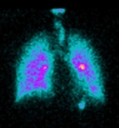
In Southampton, a research is being carried out based on advanced 3D imaging that could enhance the lives of patients with chronic lung disease.
 Southampton research uses 3D imaging to improve the lives of lung disease patients
Guided by the Professor of Inhalation Sciences at the University of Southampton, Joy Conway, the research aims to offer a better insight about various diseases such as cystic fibrosis, asthma and chronic bronchitis.
With the help of a gamma camera and CAT scanner, the 3D imaging identifies the disease and also the effect of the drugs administered within a patient's lungs. By integrating the data acquired from these two specialized medical equipment, a unique 'map' is plotted.
Data interpretation is provided by the Southampton physicists. A distinct 360º image is then generated from the scans of volunteer patients that illustrate how a specific drug is inhaled, scattered and exhaled from the patents’ lungs.
With the help of the image, the potential to regulate the infusion and delivery of inhaled drugs can be determined, in addition to developing gene therapies for diseases like cystic fibrosis. Furthermore, it will enable physiotherapists to offer better chest physiotherapy that eliminates lung secretions.
With multiple sponsorships, the latest project intends demonstrating and determining chronic obstructive pulmonary disease (COPD). It forms the major part of clinical trials and computational research carried out by the imaging team, whose findings will enhance patient care in the broader NHS.
The multidisciplinary research team at Southampton General Hospital includes university experts such as physicists and scientists as well as clinicians, nuclear medicine experts, pharmacists and radiographers.