 By Ankit SinghReviewed by Susha Cheriyedath, M.Sc.Jul 22 2024
Sexually transmitted infections (STIs) represent a critical public health concern. According to the World Health Organization (WHO), more than one million STIs are acquired every day worldwide, with the Centers for Disease Control and Prevention (CDC) reporting notable increases among young people aged 15-24 years.
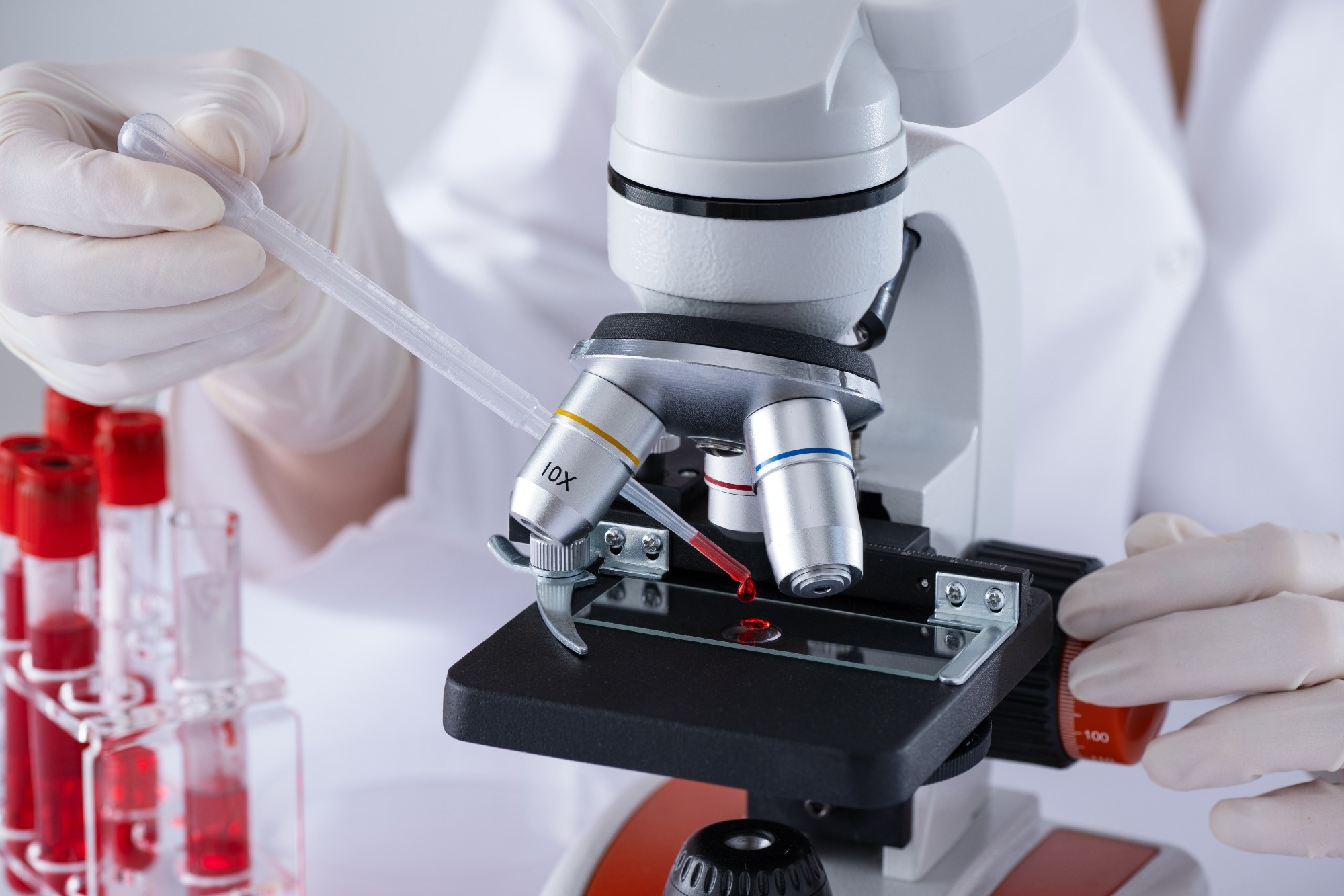
Image Credit: Inna Dodor/Shutterstock.com
Early and accurate detection is crucial in managing STIs and preventing their spread. Microscopy is a primary tool in this diagnostic process. This article explores the principles of microscopy and how advanced techniques are employed to detect STIs.
From Van Leeuwenhoek to Modern Microscopy
Microscopy has come a long way since its invention in the late 16th century by Antonie van Leeuwenhoek. His simple, single-lens microscopes allowed the first glimpses of bacteria, laying the groundwork for modern microbiology.
Key advancements include the compound microscope in the 17th century, electron microscopy in the 1930s, and recent innovations in confocal and super-resolution microscopy.1
Microscopy uses lenses to magnify small, visually imperceptible objects or structures. Light microscopy utilizes visible light and a lens system to enlarge specimens. Electron microscopy, including transmission and scanning variants, uses electron beams to achieve significantly higher resolutions. More contemporary approaches, such as confocal microscopy, use laser illumination to scan samples, generating highly detailed, three-dimensional images.1
Fluorescence Microscopy in Chlamydia Detection
Chlamydia trachomatis, the bacterium causing chlamydia, is a major cause of STIs globally. Fluorescence microscopy plays a critical role in its detection. This involves staining the bacteria with dyes that fluoresce upon binding to specific cellular components, making the bacteria appear dark against a bright background under the microscope.4
Fluorescence microscopy is highly sensitive and can detect very low levels of bacteria in clinical samples. Studies have shown that combining fluorescence microscopy with other diagnostic techniques can enhance the accuracy and speed of chlamydia detection, making it a valuable tool in clinical settings.
Detecting Gonorrhea with Gram Staining
Neisseria gonorrhoeae, the bacterium responsible for gonorrhea, can be detected using Gram staining, a classical microscopy technique. This method involves staining clinical samples with crystal violet and iodine, washing with alcohol, and counterstaining with safranin.
Neisseria gonorrhoeae appears as Gram-negative diplococci (red-colored) under the microscope, typically found within white blood cells. Gram staining is a rapid and cost-effective method widely used in clinical laboratories for the initial diagnosis of gonorrhea.5
The technique's effectiveness is enhanced when combined with culture methods, providing both immediate and confirmatory results. Studies have shown that digital imaging systems can further improve the accuracy and reproducibility of Gram stain interpretations, aiding in more reliable diagnoses.
Syphilis Detection with Dark-Field Microscopy
Syphilis, caused by the bacterium Treponema pallidum, can be detected using dark-field microscopy, which helps visualize the spirochetes in primary and secondary syphilis lesions.
In dark-field microscopy, the specimen is illuminated from the side, making the background appear dark, with the spirochetes bright due to their reflective nature. This technique also enables the visualization of the bacteria in the clinical samples, providing a definitive diagnosis.
Recent advancements, including digital enhancements and automated systems, have improved detection sensitivity and user experience.6
Electron Microscopy for High-Resolution HPV Detection
Human papillomavirus (HPV), a significant cause of cervical cancer, is extremely small and challenging to detect. Electron microscopy enables direct visualization of viral particles. This technique involves the preparation of thin sections of infected tissue and examining them under a scanning electron microscope (SEM).7
Though resource-intensive and time-consuming, electron microscopy offers detailed imagery of viral particles, confirming the presence of HPV.
Recent progress in sample processing and imaging has improved the effectiveness of electron microscopy in visualizing HPV, making it a useful tool in the investigation of the virus’s morphology and several phases in its life cycle.
Confocal Microscopy: Examining Herpes Simplex Virus
Herpes simplex virus (HSV) causes genital herpes, a common STI. Confocal microscopy is increasingly used for detailed study of HSV infections. This technique employs laser illumination to scan samples, generating high-resolution, three-dimensional images. By incorporating fluorescent antibodies that target HSV proteins, confocal microscopy enables precise localization and quantification of the virus within infected cells.8
This methodology is significant for diagnostic applications and research, offering insights into the virus's behavior and interactions with host cells. Advances in confocal microscopy, including the development of super-resolution techniques, have further improved the ability to study HSV at a molecular level, enhancing the understanding of viral pathogenesis and treatment responses.
Trichomoniasis Detection with Phase-Contrast Microscopy
Trichomoniasis, caused by the protozoan parasite Trichomonas vaginalis, is another common STI. Phase-contrast microscopy effectively detects this parasite, enhancing the contrast of transparent specimens without staining. Trichomonas vaginalis appears as motile, flagellated organisms under the microscope.9
Phase-contrast microscopy enables the identification of this parasite in vaginal swabs and other clinical specimens. This technique is particularly valuable for quick, point-of-care diagnostics, providing prompt results to guide treatment. Studies have demonstrated the high sensitivity and specificity of phase-contrast microscopy in detecting Trichomonas vaginalis, reinforcing its value in clinical practice.
Cutting-Edge Microscopy in STI Detection
Recent studies have expanded the application of microscopy in detecting STIs, utilizing cutting-edge technologies and methods to enhance diagnostic accuracy and provide deeper insights into the pathogens involved.
A recent Nature Communications study used super-resolution microscopy to investigate the interaction between Chlamydia trachomatis and host cells. Researchers used structured illumination microscopy (SIM) to visualize infection stages at a resolution beyond conventional light microscopy, revealing details about pathogen entry mechanisms and intracellular behavior.
This study highlights the potential of advanced microscopy techniques to uncover novel aspects of STI pathogenesis, contributing to the development of targeted therapies.10
In another study in Genes, researchers utilized fluorescence in situ hybridization (FISH) and confocal microscopy to detect HPV in cervical tumors. This approach enabled the specific localization of viral RNA within infected cells, providing precise diagnostic information. The study demonstrated that FISH combined with confocal microscopy could improve the sensitivity and specificity of HPV detection compared to traditional methods, making it a powerful tool for early diagnosis and monitoring of cervical cancer risk.11
The Future of Microscopy in STI Detection
Microscopy continues to evolve, with new techniques and technologies improving STI detection and study. Advances in digital and computational microscopy, such as AI-assisted image analysis, promise to enhance diagnostic accuracy and efficiency. These innovations could lead to faster, more reliable STI testing, benefiting public health worldwide.
In conclusion, microscopy remains a cornerstone in STI detection and study. From classical methods like Gram staining and dark-field microscopy to advanced techniques like confocal and electron microscopy, each approach offers unique advantages in diagnosing different STIs. As technology progresses, integrating novel imaging techniques will further enhance the ability to combat these infections, improving patient outcomes and reducing the global burden of STIs.
More from AZoOptics: Innovations in Coating Evaluation Using Impedance Spectroscopy
References and Further Reading
- Andreas P. et al. (2022). Live cell microscopy: From image to insight. Biophysics Rev. DOI: 10.1063/5.0082799
- World Health Organization (WHO). (2024). Sexually transmitted infections (STIs). [Online] World Health Organization (WHO). Available at: https://www.who.int/news-room/fact-sheets/detail/sexually-transmitted-infections-(stis)
- Centers for Disease Control and Prevention (CDC). (2024). Sexually Transmitted Infections Surveillance. [Online] Centers for Disease Control and Prevention (CDC). Avaialble at: https://www.cdc.gov/std/statistics/2022/overview.htm
- Lurie, M., et al. (2022). A Comparison of Fluorescent Microscopy Methods for the Detection of Chlamydia trachomatis. University of Cape Town. DOI: 10.25375/uct.16866583.v1
- Lachyan, A., et al. (2023). Comparison of Microscopy, Culture and Molecular Methods for Diagnosing Gonorrhea. International STD Research & Reviews. DOI: 10.9734/isrr/2023/v12i2162
- Luo, Y., Xie, Y., Xiao, Y. (2021). Laboratory Diagnostic Tools for Syphilis: Current Status and Future Prospects. Frontiers in Cellular and Infection Microbiology. DOI: 10.3389/fcimb.2020.574806
- Itoh, T., et al. (2023). Identifying Active Progeny Virus Particles in Formalin-Fixed, Paraffin-Embedded Sections Using Correlative Light and Scanning Electron Microscopy. Laboratory Investigation. DOI: 10.1016/j.labinv.2022.100020
- Nath, P., Kabir, MA., Doust, SK., Ray, A. (2021). Diagnosis of Herpes Simplex Virus: Laboratory and Point-of-Care Techniques. Infectious Disease Reports. DOI: 10.3390/idr13020049
- Li, L., Liu, J., Wang, S., Wang, X., Xiang, TZ. (2022). Trichomonas Vaginalis Segmentation in Microscope Images. Medical Image Computing and Computer Assisted Intervention, Lecture Notes in Computer Science. DOI:10.1007/978-3-031-16440-8_7
- Götz, R., et al. (2020) Nanoscale imaging of bacterial infections by sphingolipid expansion microscopy. Nat Commun. DOI: 10.1038/s41467-020-19897-1
- Mungenast, F., et al. (2021). Next-Generation Digital Histopathology of the Tumor Microenvironment. Genes. DOI: 10.3390/genes12040538
Disclaimer: The views expressed here are those of the author expressed in their private capacity and do not necessarily represent the views of AZoM.com Limited T/A AZoNetwork the owner and operator of this website. This disclaimer forms part of the Terms and conditions of use of this website.