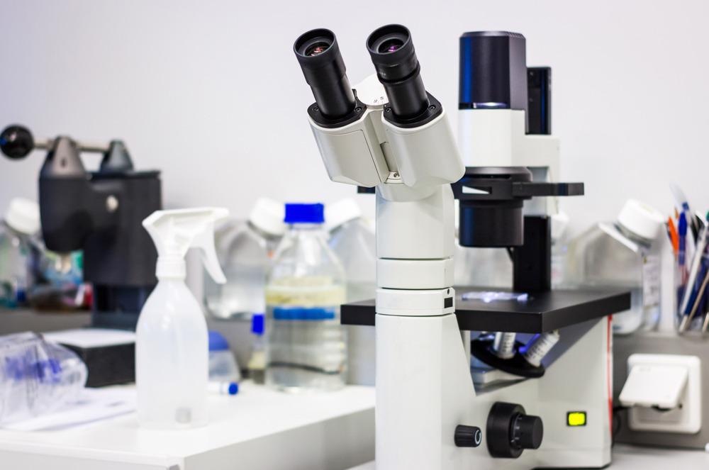
Phase-contrast microscopy transmits light through a sample. However, rather than simply looking at the relative intensity attenuation of the sample’s light, as would be done for brightfield microscopy techniques, phase contrast microscopy relies on the visualization of phase shifts in the electromagnetic light field to reveal structural information.

Image Credit: Catalin Rusnac/Shutterstock.com
With phase-contrast microscopy, it is possible to reveal cellular structures that would be invisible in a bright field experiment and would require the use of stains and often kills the cells. Phase-contrast microscopy uses a reference beam, which does not pass through the sample, and the main beam which does.
As different parts of the sample have different thicknesses and refractive indices, the main beam will undergo diffraction and scattering events. This results in a phase shift of the light that can be detected by interfering with the unshifted, reference beam.
Due to the constructive or destructive interference of the two sets of light waves, regions of this combined interference pattern will appear darker or brighter than the unperturbed reference. Different regions of the sample will also induce a different amount of phase shift on the incident light and therefore result in variation in the intensity modulation of the resulting interference pattern. Such changes can be seen by the eye or captured using a standard camera.
Phase Contrast Microscope
There are many similarities between the phase-contrast microscope and standard optical microscope design. The microscope consists of a light source, a phase condenser – to create a narrow, hollow cone of light for illuminating the sample - and a diffraction plate. The diffraction plate is located at the back focal plane of the objective lens and helps to reduce the intensity of the incident light. This is because the diffracted reference beam can be significantly brighter than the beams diffracted by the specimen.1
The choice of the annulus in the condenser needs to match the magnification and numerical aperture of the microscope in question.
Developments in Phase Contrast Microscopy
Both positive and negative phase contrasts can be used to visualize samples. In a positive phase contrast experiment, the objects with refractive indices higher than the background form dark structures with a bright halo surrounded by a second darker halo. The reverse is the case for negative phase contrast experiments.
The lack of need for sample preparation such as staining has made phase-contrast microscopy a powerful tool for investigating live cells as it eliminates the issues with phototoxicity and photobleaching found in fluorescence microscopy techniques.2 As fluorescence labels must bind and interact with the sample of interest, there is also some concern that this can result in distortion or modification of cell environments. Therefore, phase contrast microscopy can record images of cells in environments close to physiological conditions.
Quantitative versions have recently been established and developments in imaging technologies have increased the spatial resolution by a factor of two.2 However, standard implementations of the technique are still limited by the numerical aperture of the objective lens used. Super-resolution versions of the method based on holographic and other non-linear approaches are possible3 and computational approaches have also been used to enhance the effective resolution of the method.4
Phase-contrast microscopy techniques have also been widely used in the X-ray regime, not just using visible light. Highly coherent synchrotron radiation sources are well-suited to microscopy methods and the shorter wavelengths used lead to an improved spatial resolution in comparison to optical implementations.5
Applications of Phase Contrast Microscopy
The main application for phase contrast microscopy has been for label-free, live-cell imaging. As widefield images can also be obtained with the method, the technique can be used to look at extended cell networks and populations. It has the advantage of being relatively inexpensive and can be used to monitor cell cultures.
The technique is also compatible with live microorganisms, including protozoa and bacteria, and, with sufficient spatial resolution, can be used to look at sub-cellular structures such as nuclei and organelles.
The halo artifact is inherent to phase-contrast microscopy but recent developments in both image processing and in the inclusion of an interferometer in the microscope design have made it possible to reduce such artifacts.5
Phase-contrast microscopy is not limited to biological samples. This method can be used to visualize surface structures in materials science, such as for the investigation of the quality of dental implants.7 The advantage of the method is that it can be used for transparent materials that could not be seen with standard microscopy methods and can be used for a range of imaging types, from the recovery of 2D to potentially 4D structures (time-resolved 3D imaging).
References and Further Reading
- Sanderson, J. B. (2021). Phase Contrast Microscopy. Encyclopedia of Life Sciences (pp. 1–4) https://doi.org/10.1038/npg.els.0002635
- Park, Y., Depeursinge, C., & Popescu, G. (2018). Quantitative phase imaging in biomedicine. Nature Photonics, 12, 578–589. https://doi.org/10.1038/s41566-018-0253-x
- Lin, Q., Wang, D., Wang, Y., Guo, S., Panezai, S., Ouyang, L., ... & Zhao, J. (2017). Super-resolution quantitative phase-contrast imaging by microsphere-based digital holographic microscopy. Optical Engineering, 56(3), 034116. https://doi.org/10.1117/1.OE.56.3.034116
- Frangi, A. F., Schnabel, J. A., Alberola-López, C., Davatzikos, C., & Fichtinger, G. (2018). Medical Image Computing and Computer Assisted Intervention – MICCAI 2018. https://doi.org/10.1007/978-3-030-00934-2
- Snigirev, A., Snigireva, I., Kohn, V., Kuznetsov, S., & Schelokov, I. (1995). On the possibilities of x-ray phase contrast microimaging by coherent high-energy synchrotron radiation. Review of Scientific Instruments, 12, 5486. https://doi.org/10.1063/1.1146073
- Nguyen, T. H., Kandel, M., Shakir, H. M., Best-popescu, C., Arikkath, J., Do, M. N., & Popescu, G. (2017). Halo-free Phase Contrast Microscopy. Scientific Reports, 7, 44034. https://doi.org/10.1038/srep44034
- Caccianiga, G., Rey, G., Caccianiga, P., Leonida, A., Baldoni, M., Baldoni, A., & Ceraulo, S. (2021). Rough Dental Implant Surfaces and Peri-Implantitis: Role of Phase-Contrast Microscopy, Laser Protocols, and Modified Home Oral Hygiene in Maintenance. A 10-Year Retrospective Study. Applied Sciences, 11, 4985. https://doi.org/10.3390/app11114985
Disclaimer: The views expressed here are those of the author expressed in their private capacity and do not necessarily represent the views of AZoM.com Limited T/A AZoNetwork the owner and operator of this website. This disclaimer forms part of the Terms and conditions of use of this website.