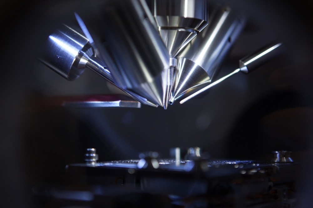
The fusion of mass spectrometry and photonics, once seemingly unrelated, has yielded a powerful toolkit that transcends previous limits on analytical science. This dynamic alliance finds applications across diverse fields such as pharmaceuticals, fuels, metabolites, food science, environmental monitoring, forensics, and beyond, unlocking new frontiers in scientific exploration.

Image Credit: Intothelight Photography/Shutterstock.com
How Do the Mass Spectrometry and Photonics Technologies Enhance One Another?
Mass spectrometry (MS) is an analytical technique that separates ions based on mass-to-charge ratio, offering detailed insights into molecular composition. In contrast, photonics is the study and application of manipulating photons, fundamental particles of light, through transmission, emission, and modulation.
The synergy between mass spectrometry and photonics enhances analytical capabilities by combining the precision of MS with the selective and rapid ionization capabilities of laser-based photonics techniques, enhancing sensitivity and enabling diverse, high-throughput analyses. It enables spatially resolved imaging, real-time monitoring, and novel ionization sources, expanding proteomics, metabolomics, environmental analysis, and materials science applications.
Their fusion has given rise to a host of innovative techniques like laser ablation and matrix-assisted laser desorption/ionization (MALDI), revolutionizing elemental analysis, tissue imaging, and biomolecule detection while also pushing the analytical boundaries with advanced ionization sources like laser-induced breakdown spectroscopy (LIBS) and photoionization.
Mass spectrometry and photonics form a powerful alliance, advancing scientific exploration and understanding of complex molecular systems.
How Is Mass Spectrometry Being Used with Photonics in Modern Research?
Illuminating In Vivo Metabolomics with MALDI Imaging
One particularly impactful synergy can be seen in metabolomics and molecular histology, with the rise of matrix-assisted laser desorption/ionization (MALDI) mass spectral imaging.
This technique entails coating tissue sections with an energy-absorbing chemical matrix before using a pulsed UV laser to systematically ablate and ionize material across the sample. The generated ions are then analyzed through time-of-flight mass spectrometry, providing high spatial resolution molecular mass and distribution data.
Compared to traditional mass spectrometry methods that require extensive sample preparation and pooling, MALDI enables the direct analysis of metabolites and lipids in intact tissue while preserving crucial histological context. Already, MALDI imaging has provided groundbreaking insight into endogenous compound distribution variances underlying diseases like cancer.
For instance, Kyushu University researchers employed matrix-assisted laser desorption ionization-mass spectrometry (MALDI-MS) for rapid, high-throughput metabolic profiling of human leukemia cells during drug exposure. By screening over 2,500-minute cell cultures in multiwell plates using MALDI time-of-flight instruments, the team could map over 150 metabolite peak responses within 90 seconds of matrix-laser ablation. This demonstrated the exciting potential of the approach for expediting pharmaceutical testing through direct drug-metabolome interaction assessments.
Similarly, advanced MALDI setups at institutions like the University of California, San Diego, have visualized the signaling networks of bacteria with subcellular. These setups captured simultaneous metabolic snapshots, illustrating the communication between these individual single-celled organisms.
These studies highlight the unprecedented glimpses that the fusion of photonics and mass spectrometry can provide into the real-time biochemistry of cells in natural environments.
Elemental Imaging of Tissues Via Laser Ablation ICP-MS
MALDI imaging is not the only mass spectrometry-photonics hybrid technique making waves in analytical science. Laser ablation inductively coupled plasma mass spectrometry (LA-ICP-MS) uses high-energy laser pulses to directly sample solid materials, with the aerosolized compounds subsequently ionized by an argon plasma source and fed into a mass spectrometer. It facilitates rapid elemental mapping across tissue and cell samples while requiring minimal sample preparation compared to traditional ICP-MS.
LA-ICP-MS is seeing growing use among neuroscientists seeking to elucidate the elemental underpinnings of neurological disorders and processes at the cellular scale. For example, researchers at Forschungszentrum Jülich in Germany employed LA-ICP-MS to construct distribution maps for copper, iron, zinc, and manganese across sections of Parkinson’s disease-affected mouse brain tissue.
Intriguing copper accumulation patterns were noted around damaged neuron sites, providing clues into the role of metal dyshomeostasis in Parkinson’s pathogenesis. This study exemplifies the new microanalytical dimensions accessed by combining laser sampling and ICP-MS capabilities.
Smaller Footprint Field Instruments
Increasing portability further expands the utility of dual mass spec and photonics devices. Compact field units are ideal for applications like forensic surveillance, food quality monitoring, and environmental threat assessment, which formerly required bulky instrumentation.
In a study published in the International Journal of Mass Spectrometry, researchers developed a portable device employing a single-photon ionization time-of-flight mass spectrometer (SPI-Tof-MS). It ionized samples using 118-nm pulses from a frequency-tripled Nd:YAG laser. Operating in reflectron Tof-MS mode, the device achieved a mass resolution of 800 at about 100 atomic mass units, demonstrating its capability for online monitoring of diesel engine exhaust.
The compact and laser-based SPI-Tof-MS offered a detection limit of parts per billion by volume (ppbv) for trace analysis of organic compounds, showcasing its potential for applications such as environmental air monitoring and automobile exhaust gas analysis.
Characterizing Material Defects & Purity with Mass Spectrometry
While the above applications focus on leveraging photonic systems for enhanced mass analyses, spectrometric techniques are also coming to play an increasingly pivotal role in photonics research itself.
Of particular note is the emergence of mass spectrometry for optical material and component characterization. Techniques like secondary ion mass spectrometry (SIMS) are standard for analyzing doping profiles and free carrier dynamics in semiconductors used in lasers, LEDs, and photovoltaics to shed light on device efficiency droops. This has been crucial for defects and interface studies related to applications like GaN high-electron mobility transistors (HEMTs).
Meanwhile, for glass and crystal growth, mass spectrometry enables researchers to identify contaminants and quantify impurity distributions that contribute to optical losses in solid-state laser gain media. Mass spectrometry has also facilitated tighter compositional control over alloyed semiconductor compounds like AlGaAs used in diode pump lasers for telecom networks - helping curb performance variability.
Challenges and Future Prospects
While the fusion of mass spectrometry and photonics has yielded remarkable advancements, there are challenges that researchers continue to address. One such challenge is the optimization of laser parameters for different sample types and analytical goals. However, continued developments in laser technology and mass analyzers are expected to overcome these challenges and further enhance the capabilities of hybrid integrated mass spectrometry and photonics systems.
More from AZoOptics: High-Precision Optical Metrology Techniques and Their Principles
References and Further Reading
Van de Plas, R., Yang, J., Spraggins, J., & Caprioli, R. M. (2015). Image fusion of mass spectrometry and microscopy: a multimodality paradigm for molecular tissue mapping. Nature Methods, 12(4), 366-372. https://doi.org/10.1038/nmeth.3296
Nellessen, C. M., & Nehl, D. B. (2023). An easy adjustment of instrument settings (‘Peak MALDI’) improves identification of organisms by MALDI-ToF mass spectrometry. Scientific Reports, 13(1), 15018. https://doi.org/10.1038/s41598-023-42328-2
Miura, D., Fujimura, Y., Tachibana, H., & Wariishi, H. (2010). Highly sensitive matrix-assisted laser desorption ionization-mass spectrometry for high-throughput metabolic profiling. Analytical chemistry, 82(2), 498-504. https://doi.org/10.1021/ac901083a
Yang, Y. L., Xu, Y., Straight, P., & Dorrestein, P. C. (2009). Translating metabolic exchange with imaging mass spectrometry. Nature Chemical Biology, 5(12), 885-887. https://doi.org/10.1038/nchembio.252
Matusch, A., Depboylu, C., Palm, C., Wu, B., Höglinger, G. U., Schäfer, M. K. H., & Becker, J. S. (2011). Cerebral bioimaging of Cu, Fe, Zn, and Mn in the MPTP mouse model of Parkinson’s disease using laser ablation inductively coupled plasma mass spectrometry (LA-ICP-MS). Journal of the American Society for Mass Spectrometry, 21, 161-171. https://doi.org/10.1016/j.jasms.2009.09.022
Tonokura, K., Kanno, N., Yamamoto, Y., & Yamada, H. (2010). Development of a compact laser-based single photon ionization time-of-flight mass spectrometer. International Journal of Mass Spectrometry, 290(1), 9-13. https://doi.org/10.1016/j.ijms.2009.11.004
Disclaimer: The views expressed here are those of the author expressed in their private capacity and do not necessarily represent the views of AZoM.com Limited T/A AZoNetwork the owner and operator of this website. This disclaimer forms part of the Terms and conditions of use of this website.