Electron microscopes are incredibly powerful analytical tools that are used to reveal and characterize the molecular structure of organic and inorganic substances and materials. This article will look at how electron microscopy is helping scientists understand an enigmatic material: Roman concrete.

Image Credit: Steve Lovegrove/Shutterstock.com
Electron Microscopy: An Overview
Electron microscopes provide researchers with more powerful imaging capabilities than conventional optical microscopes due to their higher resolving power. Optical microscopes are limited by the wavelength of visible light and typically only resolve objects over 200 nm.
Transmission electron microscopes and scanning electron microscopes can magnify a sample up to 10,000 x, whereas optical microscopes can only provide a maximum useful resolution of 2000 x. Electron microscopy is used to reveal the intimate microstructural details of crystals, cells, microorganisms, metals, and many other inorganic materials.
The Mysteries of Roman Concrete
The modern world has been built with concrete. This cheap, abundant material provides civil engineers and planners with the ability to construct domestic dwellings, commercial buildings, and infrastructure and meet the demands of a rapidly growing global population.
Modern concrete, however, has limited durability. Concrete structures can start to experience problems within just a few decades of being built. Concrete is especially vulnerable to decay in marine environments.
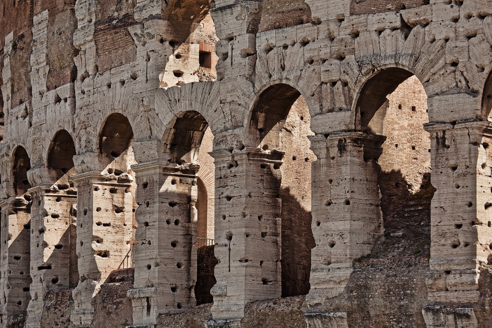
Many people might think of concrete as a uniquely modern building material, but the Romans were using it to construct public buildings around two thousand years ago. Amongst the many impressive concrete structures which are still standing is the Pantheon, which contains the world’s largest unreinforced concrete dome.

Image Credit: S.Borisov/Shutterstock.com
Romans used a unique type of volcanic ash in their concrete, known as Pozzolana, which reacts at room temperature with calcium hydroxide in the presence of water. Additionally, enhanced binding performance is enhanced in Roman concrete by the presence of highly porous glass.
The use of lime in concrete also helps the strength of the material increase over time. This is especially pronounced in the presence of seawater. Until recently, the contribution of lime to the durability of Roman concrete was largely overlooked by researchers.
Electron Microscopy Reveals the Secrets of Roman Concrete’s Longevity
Analysis of Roman concrete using techniques such as electron microscopy has revealed the presence of lime clasts, which are millimeter-sized pieces of lime. For a long time, these clasts were thought to be the result of low-quality control by Roman builders. However, recent research by scientists from MIT has revealed their key contribution.
Researchers have analyzed 2,000-year-old samples of Roman concrete from an archaeological site near Rome, using advanced electron microscopy and X-ray diffraction techniques to study the material’s composition and structure.
Roman builders used a process called hot mixing, which adds the lime at a high temperature without incorporating water. Under elevated temperatures, clasts with brittle nanosized architectures are produced, which function as a calcium source that provides concrete with self-healing capabilities.
Experimental phases in the research were as follows: specimen preparation, crack validation, specimen preconditioning, and crack healing. Continuous water flow over a 30-day period revealed a measured decrease in the flow rate of samples containing lime clasts, revealing potential self-healing activity in the samples of Roman concrete.
How Was Electron Microscopy Utilized in this Research?
The researchers behind the study of lime clasts in Roman concrete utilized quantitative SEM-EDS. Both polished and fractured samples were analyzed in the study. Element analysis was utilized to study the spatial distribution and composition of phases, especially the lime clasts and surrounding matrix in the mortar.
The chemistry of the freshly fractured and polished samples was also analyzed in detail by MIT scientists. Fractured samples were used to avoid any chemical alteration of clasts during polishing techniques. Samples were imaged in low vacuum conditions. Datasets were divided into clusters using fuzzy clustering techniques.
Why Electron Microscopy is Suited to This Research
Electron microscopy provides significantly enhanced capabilities for the study of concrete petrography. The microstructure of cement, concrete, and aggregates is revealed in unprecedented detail by the enhanced spatial resolution and contrast provided by electron microscopy techniques.
As well as providing detailed information on the microstructural characteristics of concrete, electron microscopy techniques such as SEM helps researchers investigate how concrete deteriorates. Quantitative information such as phase abundance, composition, and phase distribution can be extracted from obtained data.
The sub-micrometer definition is provided by SEM techniques, but researchers must be aware of the need for careful and proper sample preparation as imaging techniques require highly polished surfaces. However, accurate segmentation of images into constituent phases can be achieved by combining X-ray and backscattered electron information.
Revealing the secrets of Roman concrete and its extreme durability requires advanced analytical and imaging techniques. Scanning electron microscopy is uniquely suited to revealing the sub-micrometer characterization of microstructural elements such as lime clasts and their contribution to the superior self-healing performance of this type of ancient building material.
In Summary
Whilst the MIT scientist’s research provides key evidence for the contribution of lime clasts, it must be noted that the Romans used different techniques and material variants to produce concrete to provide its unique properties.
Using electron microscopy and other advanced analytical techniques, the secrets of Roman concrete are being revealed. Current research in this field of material science could provide new and intriguing insights into improving modern concrete for applications such as sustainable, self-healing 3D-printed concrete.
More from AZoOptics: 4D Scanning Transmission Electron Microscopy (4D-STEM) for Battery Analysis
References and Further Reading
Andrei, M (2023) Researchers uncover the secrets behind the insane durability of Roman concrete [online] zmscience.com. Available at: https://www.zmescience.com/science/researchers-uncover-the-secrets-behind-the-insane-durability-of-roman-concrete/
Seymour, L.M et al. (2023) Hot mixing: Mechanistic insights into the durability of ancient Roman concrete Science Advances 9(1) [online] science.org. Available at: https://www.science.org/doi/10.1126/sciadv.add1602
Stutzman, P.E (2000) Scanning Electron Microscopy in Concrete Petrography [online] nist.gov. Available at: https://www.nist.gov/publications/scanning-electron-microscopy-concrete-petrograph
Disclaimer: The views expressed here are those of the author expressed in their private capacity and do not necessarily represent the views of AZoM.com Limited T/A AZoNetwork the owner and operator of this website. This disclaimer forms part of the Terms and conditions of use of this website.