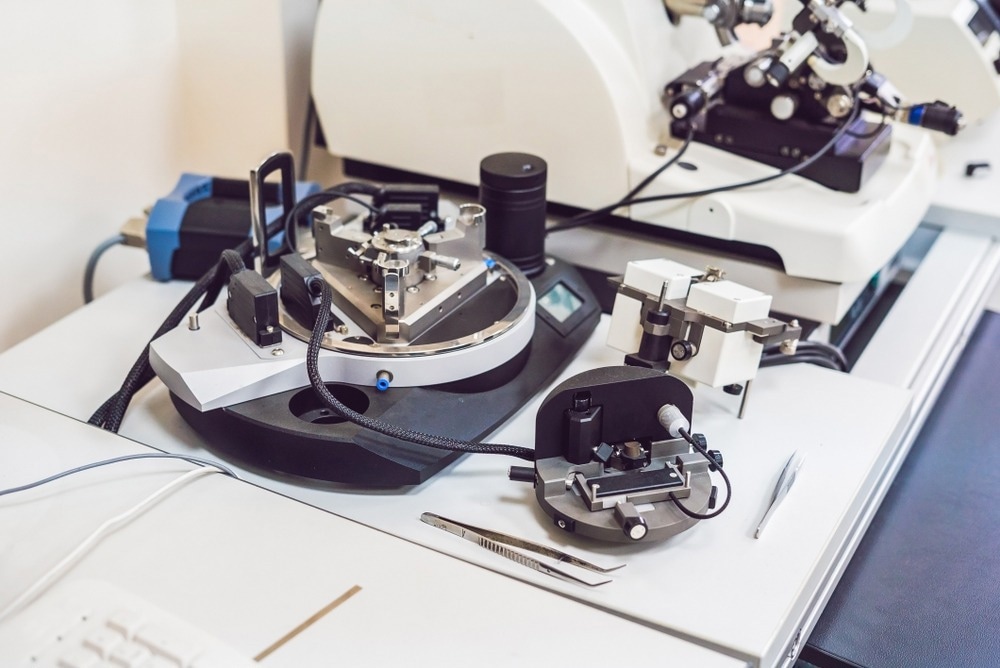
Atomic force microscopy (AFM), a form of scanning probe microscopy (SPM), exhibits resolution on the order of fractions of a nanometer. The data is obtained by sensing or touching the surface with a mechanical probe. Piezoelectric components allow for small, exact movements under electrical control, enabling impeccable scanning. The atomic force microscope does not employ nuclear force, despite its name.

Image Credit: Elizaveta Galitckaia/Shutterstock.com
Features in Atomic Force Microscopy
The three main capabilities of the AFM are force measurement, topography imaging, and manipulation.
The forces between the probe and the sample as a function of their mutual separation can be measured using AFMs in force measurement. This can be used to do force spectroscopy and evaluate the sample's mechanical characteristics, such as its Young's modulus.
For imaging, it is possible to create a high-resolution image of a sample surface's three-dimensional shape (topography) using the probe's response to the forces the sample exerts on it. This is accomplished by raster scanning. The position of the sample concerning the tip and logging the height of the probe corresponds to a steady interaction between the probe and the sample. A pseudo color plot is a common way to show the surface topography.
Working Principle of AFM
The AFM consists of a cantilever with a probe-like sharp tip at one end that scans the object's surface. The cantilever is commonly made of silicon or silicon nitride with a tip radius of curvature on the order of nanometers. Hooke's law states that when the tip is brought close to a sample surface, forces between the tip and the sample cause the cantilever to deflect. Mechanical contact forces, capillary forces, van der Waals forces, and chemical bonding are among the forces that can be measured in AFM depending on the circumstance. It is possible to measure multiple quantities simultaneously using specific probes.
Various operating modes for the AFM are available depending on the application. The various dynamic (non-contact or "tapping") imaging modes, in which the cantilever oscillates or vibrates at a specific frequency, are split into two categories: static (also known as a contact) modes and dynamic (non-contact) modes in general.
Applications of Atomic Force Microscopy
AFM can be used for many natural scientific disciplines, including solid-state physics, the energy sector, and semiconductor technology. Studying changes in physical properties resulting from atomic manipulation is one application of solid-state physics. Other applications include the identification of atoms at a surface, the assessment of interactions between a particular atom and its neighboring atoms, and evaluating changes in physical properties resulting from atomic manipulation.
Atomic Force Microscopy Used for Energy Research
Atomic force microscopy is a decent candidate as a characterization technique for energy research because it can locally examine nanoscale electrochemical processes. The power density and lifespan of next-generation materials used in energy storage systems such as lithium-ion batteries, fuel cells, and supercapacitors are being extended by applying various AFM methods. AFM can also be used to investigate local ionic transport and reactivity.
The development of renewable energy storage techniques is critically needed to satisfy the performance needs of our society. The basic knowledge of the electrocatalytic challenges is the requirement for battery improvements. AFM offers high-resolution morphological information at each interface within the electrochemical systems. In situ, AFM experiments demonstrate real-time imaging of interfacial reactions at the nanoscale.
Over the last two decades, atomic force microscopy (AFM)-based methods have played a significant role in understanding the structure and characteristics of photovoltaic materials with high temporal and spatial resolutions. Simultaneously, the sample morphological characters can be connected to their electrical, chemical, and photonic properties, which points out the linkage between the performance and effectiveness of these complex devices. AFM has become a vital method for multiparametric and multipurpose characterizations of solar cells. The photovoltaics evaluation methods include photoconductive AFM (pc-AFM), time-resolved electric force microscopy, and Kelvin probe force microscopy (KPFM).
Advantages of Atomic Force Microscopy
AFM is superior to other microscopes in several ways. The AFM offers a three-dimensional surface profile as opposed to a sample's two-dimensional projection or image of an electron microscope.
AFM samples do not suffer from charging in the final image and do not require any specific treatments (such as metal or carbon coatings) that can permanently alter or affect the sample.
AFM can function accurately in ambient air or even a liquid environment; other microscopes require an expensive vacuum environment for effective functioning. Studying biological macromolecules and even living things is now possible as a result. AFM can theoretically offer more resolution.
Limitations of Atomic Force Microscopy
The single scan image size is a drawback of AFM. The AFM can only image a maximum scanning area of around 150 micrometers and a maximum height on the order of 10-20 micrometers, whereas the electron microscope can image an area on the order of square millimeters with a depth of field on the scale of millimeters in one scan.
Using parallel probes such as millipede data storage is one way to increase the size of the scanned region for AFM.
Conclusion
In the context of energy research, atomic force microscopy (AFM) can be used to examine and quantify the physical characteristics of several types of materials at the nanoscale in the atmosphere, liquid phase, and ultra-high vacuum environment. It is now a crucial instrument for applications in energy research.
AFM is employed in many energy research applications, such as bioenergy, geothermal, photovoltaics, renewable fuels, and solar panels.
References
Chen, J., & Xu, K. (2020). Applications of atomic force microscopy in materials, semiconductors, polymers, and medicine: A minireview. Instrumentation Science & Technology, 48(6), 667–681. https://doi.org/10.1080/10739149.2020.1764030
Kosareva, E. K., Pivkina, A. N., & Muravyev, N. V. (2022). Atomic force microscopy in energetic materials research: A review. Energetic Materials Frontiers. https://www.sciencedirect.com/science/article/pii/S2666647222000410
Disclaimer: The views expressed here are those of the author expressed in their private capacity and do not necessarily represent the views of AZoM.com Limited T/A AZoNetwork the owner and operator of this website. This disclaimer forms part of the Terms and conditions of use of this website.