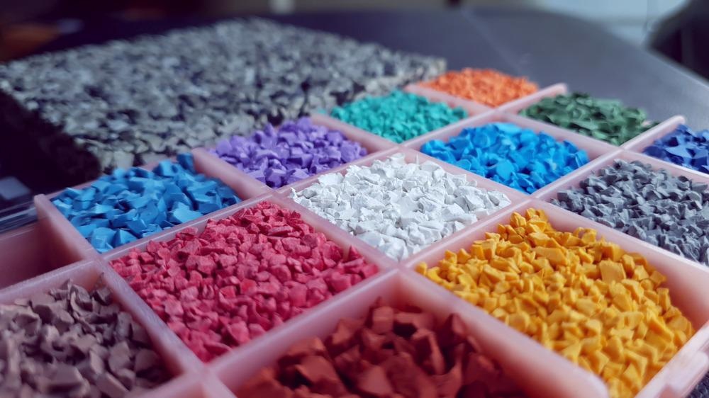
The great industrial success of the polymer materials, such as plastics, rubbers, textiles, biopolymers, and many others, led to the emergence of entirely new classes of functional materials. These novel materials require accurate characterization to gain an understanding of their complex structure and morphology and to determine their most suitable application.

Image Credit: Wongsakorn Dulyavit/Shutterstock.com
Various microscopy techniques enable multifunctional and in-depth material characterization over a wide range of length scales (ranging from millimeters down to nanometers) and can provide physical, chemical, and structural information, associated with the performance of the material.
Polymer materials have become ubiquitous in modern society with various applications, ranging from textiles, consumer goods manufacturing, and packaging, through construction, transportation, and electronics to medicine and automotive and aircraft manufacturing. These materials are strong, durable, lightweight, low cost, and have a wide range of desirable chemical properties.
What is a Polymer?
A polymer is a large organic molecule (macromolecule) consisting of smaller repeat units (called monomers) that are bound to each other with covalent bonds.
The exceptional durability and remarkable mechanical properties of the polymers result from these high-strength covalent bonds that form dense macromolecular networks. Compared to traditional materials such as metal, minerals, and wood, modern polymers offer superior versatility and mechanical properties combined with excellent stability and processability. These properties define the dominant place that polymers have in the modern economy.
Throughout the years, major developments in polymer materials science and technology resulted in advanced synthetic methods and new polymerization routes, allowing superior control of the macromolecular structure of the polymers.
In parallel, new microscopy techniques were developed for better characterization of the structure of the polymer macromolecules. This, in turn, enabled the synthesis of specialty polymers with tunable properties (achieved by tailoring the structure and arrangement of the polymer chains) that suit different applications.
Microscopy Reveals Key Structure-Function Relations in Polymers
The microscopic characterization of polymers is key to the understanding of the fundamental structure-function relation on a molecular level and paves the way toward the rational design of novel and improved polymers materials.
Many different microscopy techniques are currently used for materials characterization, with fluorescence microscopy, electron microscopy, and atomic force microscopy being the most relevant ones.
Fluorescence microscopy is an optical characterization technique that uses fluorescence (emission of photons by a substance that has absorbed electromagnetic energy) instead of (or in addition to) light scattering, reflection, and absorption.
Fluorescence microscopy offers a high signal-to-noise ratio, chemical specificity, and multiwavelength ability.
The technique is commonly used to study the kinetics of processes such as crystal growth and phase separation in polymer materials on the microscale. While diffraction limits the spatial resolution of traditional light-based microscopes to a few hundred nanometers, scientists have recently made exciting breakthroughs to bypass this limit.
Optical Super-Resolution Microscopy in Polymer Science
Following ground-breaking work on single-molecule imaging in the 1990s, several super-resolution optical microscopy methods have been developed, including stimulated emission depletion and single-molecule localization microscopy, that enabled sub-diffraction-limited spatial resolution imaging by confining the illuminated volume of the sample resulting from selective activation/deactivation and localization of fluorophores (fluorescent labeling molecules).
Such super-resolution techniques provide a spatial resolution that is one order of magnitude higher than the diffraction limit (typically several tens on nanometers).
Super-resolution optical microscopy studies have already provided significant new insights into the conformational behavior of macromolecules and interfacial dynamics of polymers during crystallization and melting.
Electron Beams Improve Imaging Resolution
Despite the advances in super-resolution microscopy, detailed structural studies of polymer materials at a molecular level (length scales of less than 20 nm) require access to even higher resolution imaging techniques. This can be achieved by employing electron beams to image the sample.
Electron microscopy extends the optical diffraction limit by using an electron beam as an illumination source. Depending on their energy, electrons have a wavelength more than 1000 times smaller compared to visible light.
Electron microscopy can be divided into transmission electron microscopy (TEM) and scanning electron microscopy (SEM). In terms of spatial resolution, SEM is somewhat intermediate between optical microscopy and TEM.
TEM allows resolution of the order of 0.1 nm or better to be achieved for inorganic crystalline samples. However, for polymer materials, the material's electron beam sensitivity and low electron density limit the achievable resolution.
A significant advantage of using SEM (compared to TEM) is the ability to image the surfaces of bulk samples with a large depth of focus.
Recent developments in cryo-electron microscopy and environmental electron microscopy permit imaging of non-stained materials in native conditions and visualization of dynamic phenomena, such as crystal nucleation and growth, with nanometer resolution.
Advanced Atomic Force Microscopy in Polymer Science
Atomic force microscopy (AFM) utilizes atomically-sharp probes for sample surface scanning. The technique offers several unique advantages, including operation at elevated temperatures (up to several hundred °C) and in a wide range of environmental conditions.
More importantly, the AFM provides atomic-level resolution, probing the conformation of individual polymer chains and the morphology of materials and coatings at an unprecedented level. However, the technique is somewhat limited by slow scan speeds.
Recent advancements in high-speed AFM significantly improved the time resolution, making it possible to visualize dynamic phenomena.
Moreover, the ability of the AFM to sense mechanical forces expands the versatility of the technique and enables scientists to characterize the mechanical and electrical properties of polymer materials.
Correlative Microscopy Combines the Strengths of Different Microscopic Techniques
Each of the techniques has its strength and weaknesses, and the different methods are highly complementary.
In recent years, this led to the development of several correlative approaches that exploit the strengths of each individual technique.
In correlative imaging, the polymer sample is characterized using two or more different microscopy techniques (for example AFM and SEM or SEM and fluorescence microscopy) and the resulting images of the same area of the sample are superposed.
This approach enables scientists to obtain more comprehensive and informative data than any of the individual techniques can offer.
Despite being in its infancy, correlative microscopy has enormous potential for material characterization.
Researchers envision that the techniques will become an important tool capable of addressing some of the most important outstanding questions in the field of polymer science.
References and Further Reading
Wang, Y., et al. (2021) Correlative imaging for polymer science. J Polym Sci, 59, 1232– 1240. Available at: https://onlinelibrary.wiley.com/doi/10.1002/pol.20210013
Michler, G.H. and Lebek, W. (2016). Electron Microscopy of Polymers. In Polymer Morphology, Q. Guo (Ed.). Available at: https://doi.org/10.1002/9781118892756.ch3
Coceancigh, H., et al. (2019) Optical Microscopic Techniques for Synthetic Polymer Characterization. Analytical Chemistry, 91 (1), 405-424. Available at: https://doi.org/10.1021/acs.analchem.8b04694
Nguyen-Tri, P., et al. (2020) Recent Applications of Advanced Atomic Force Microscopy in Polymer Science: A Review. Polymers, 12(5), 1142. Available at: https://doi.org/10.3390/polym12051142
Venkateshaiah, A., et al. (2020) Microscopic Techniques for the Analysis of Micro and Nanostructures of Biopolymers and Their Derivatives. Polymers, 12(3), 512. Available at: https://doi.org/10.3390/polym12030512
Chapman, D. V., et al. (2020) Optical Super-Resolution Microscopy in Polymer Science. Progress in Polymer Science, 101312. Available at: https://doi.org/10.1016/j.progpolymsci.2020.101312
Disclaimer: The views expressed here are those of the author expressed in their private capacity and do not necessarily represent the views of AZoM.com Limited T/A AZoNetwork the owner and operator of this website. This disclaimer forms part of the Terms and conditions of use of this website.