Chemical and physical phenomena at the interface between water and solids play a significant role in many biological processes and industrial technologies. Understanding the interactions at the water-solid interfaces at the molecular level has been challenging. However, advancements in atomic force microscopy (AFM) have enabled atomic resolution imaging of water-solid interfaces.

Image Credit: OlegRi/Shutterstock.com
Importance of Understanding Water-Solid Interfaces
Water-solid interfaces are significant due to their strong relevance to human life. For example, many biomolecules interact with water or other molecules, especially at the interfaces between physiological solutions in the human body. Since the human body mostly consists of aqueous solutions, water-sold interactions play an important role in biological processes.
At a water-solid interface, the surface exerts a force in the water molecules. This force can extend to distances as much as a few water layers. The impact of the surface on the water molecule is in all directions.
Spectroscopic techniques, such as x-ray or neutron diffraction, are typically used to image water-solid interfaces. But these methods can only provide limited information. The images are mostly constricted to the surface of the molecule. Atomic force microscopy (AFM) is a powerful imaging technique that allows probing of the water-solid interface with high resolution.
Using AFM for Imaging
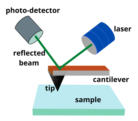
Atomic force microscopy (AFM) is a powerful microscopy technique that is widely used for high-resolution imaging. AFMs are equipped with a cantilever with a sharp tip at its end as a force sensor. The back of the cantilever is coated with a reflective “mirror” type substance and a laser beam is directed at the back of the cantilever. The reflected beam is detected by a photodetector (Figure 1).
When the tip feels a force, the cantilever oscillates up and down. The motion of the cantilever deflects the laser beam which is detected and imaged by the photo-detector. High-resolution measurements can be carried out in the vertical and lateral directions by scanning the cantilever.

Figure 1: Schematic illustrations showing basic principles of AFM. Image Credit: Ilamaran Sivarajah
Three-Dimensional Imaging with AFM
AFM allows for high-resolution imaging in three dimensions (3D). To obtain images in all three dimensions, an advanced AFM technique called frequency modulated atomic force microscope (FM AFM) is used.
FM AFM introduces frequency modulation to the AFM cantilever. The cantilever does not contact the sample but oscillates above the surface. Frequency modulation is a controlled oscillation of the tip of the cantilever. Any weak force encountered by the tip is detected by the change in the oscillation frequency.
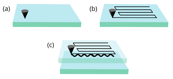
The FM AFM is scanned across the sample surface to capture images in the X, Y, and Z directions (Figure 2). From images obtained in a 3D scan, any one-dimensional (1D) or two-dimensional (2D) information can also be extracted. Using FM AFM water distribution at a water-solid interface can be investigated more efficiently and accurately than other techniques.
Visualization of 1D profiles of the hydration force, 2D images of hydration layers, and 3D distributions of water molecules have been demonstrated using FM AFM.

Figure 2: Schematic illustrations showing basic principles of (a) 1D AFM, (b) 2D AFM, (c) 3D AFM. Image Credit: Ilamaran Sivarajah
qPlus-Based AFM with Functionalized Tips
AFM using optical detection can be challenging in some experimental environments. For example, in low temperatures where laser-induced thermal effects can interfere with measurements. Various water-solid interfaces are studied at low temperatures. To overcome this limitation, German physicist Franz Josef Giessibl invented the qPlus sensor in 1996. The qPlus sensor uses a quartz tip instead of traditional silicon-based or other material-based tips. Quartz has a piezoelectric property that is well-suited for AFM tips.
The piezoelectric effect is the ability of certain materials to generate an electric charge in response to applied mechanical stress. This capability of quartz can replace the need to use a laser for detection. qPlus-based tips can be functionalized with different piezoelectric materials to suit the experiment.
qPlus-based AFMs with functionalized tips have been demonstrated to produce images of unprecedented resolution on various water-solid interfaces. 1D water chain, 2D water layers, and mixed water-hydroxyl layers, water clusters, and ion hydrates have been studied using qPlus-based AFMs.
Achievements and Improvements to AFM for High-Resolution Imaging
The advancement of AFM with new techniques such as FM-AFM and qPlus-based AFMs with functionalized tips have progressed the knowledge of water-solid interfaces. These findings have contributed to the development of new technology. They have also deepened our understanding of biological processes towards disease prevention as well as new drug discovery.
To continue the progress of AFM-based techniques, several challenges have to be overcome. The scan speed of the AFM is limited. This is due to the mechanical integration of the tip materials and the detection apparatus. Optimizing the various components of the AFM with technology developed in other fields offers viable solutions. For example, high-speed cameras and high-speed optomechanical elements increase scanning speed. Further improvement of the force sensitivity could also enhance the performance and results of the AFM. Ideas for combining other experimental techniques such as vibrational spectroscopy or adapting single photon-based methods developed in condensed matter physics have been proposed.
References and Further Reading
Franz J. Giessibl (2019) The qPlus sensor, a powerful core for the atomic force microscope. Review of Scientific Instruments, 90, 011101, https://doi.org/10.1063/1.5052264
Jinbo Peng, Jing Guo, Runze Ma, Ying Jiang (2021) Water-solid interfaces probed by high-resolution atomic force microscopy. Surface Science Reports, 100549,ISSN 0167-5729, https://doi.org/10.1016/j.surfrep.2021.100549
Takeshi Fukuma (2010) Water distribution at solid/liquid interfaces visualized by frequency modulation atomic force microscopy, Science and Technology of Advanced Materials, 11:3, https://doi.org/10.1088/1468-6996/11/3/033003
Marchesi, A., Umeda, K., Komekawa, T. et al. (2021) An ultra-wide scanner for large-area high-speed atomic force microscopy with megapixel resolution. Sci Rep 11, 13003, https://doi.org/10.1038/s41598-021-92365-y
Disclaimer: The views expressed here are those of the author expressed in their private capacity and do not necessarily represent the views of AZoM.com Limited T/A AZoNetwork the owner and operator of this website. This disclaimer forms part of the Terms and conditions of use of this website.