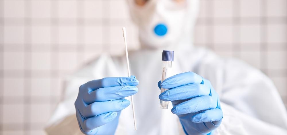
Raman spectroscopy has evolved into a powerful tool for both medical analysis and forensics.1 The ability to perform qualitative and quantitative chemical analysis on a variety of sample types has made Raman spectroscopy an excellent technique for analyzing the chemical composition of samples or looking for biomarkers as part of medical diagnosis.

Image Credit: Fusionstudio/Shutterstock.com
Raman Spectroscopy and Vibrational Modes
Raman spectroscopy is sensitive to certain vibrational modes in a molecular structure that are ‘Raman-active’. Raman-active vibrational modes induce a change in the polarizability of the molecule during the vibrational motion. For larger molecular systems, there is normally a sufficiently large number of Raman active vibrational modes that is a ‘fingerprint’ of the molecular structure. Matching these fingerprint modes to a reference spectrum in a database is a commonly used way of automated identification of chemical species with Raman spectroscopy.
Medical diagnosis often uses tissue or fluid samples from the patient. Raman imaging methods are becoming increasingly popular in histopathology or point-of-care diagnostic methods. This is for both for the detection of the biomarkers that indicate the presence of tumors but also for the investigation of how medical therapies are working and to investigate the surrounding tumor microenvironment.1,2
As Raman spectroscopy can be used to measure the chemical composition of liquids, it is inherently compatible with biofluid measurement. One challenge when analyzing the composition of biofluids is that the sample can need either pre-concentration or preparative steps before analysis. This is particularly true for samples such as blood where a significant amount of preparative work is needed before the analysis can be made.
Another consideration when choosing a biofluid type for analysis and potential diagnosis is the achievable volume. Saliva and urine are both relatively non-invasive samples to collect from a patient, unlike blood. Blood samples are very restricted in how much volume can be taken from the patient but also blood testing also requires relatively large volumes for analysis.
Saliva Samples
Ninety percent of saliva is secreted from salivary glands. It has been used successfully as a biofluid for medical diagnosis of diseases such as cancer, viral and other types of oral infection, cardiovascular disease, and diabetes.3 The success of using saliva as a biofluid for analysis and the fact that samples are easy to collect from patients and non-invasive has led to the development of salivaomics – the range of microRNA, transcriptomics, proteomics, and metabonomics analysis of saliva samples.
Raman spectroscopy has been applied for the analysis of salivatory samples for both medical diagnostics and forensics tests, for example for the screening of the presence of illegal drugs.4 Results have shown a sensitivity down to 10 µgL-1 for the presence of drugs such as cocaine. Advances in the portability of Raman spectrometers will make it possible to create robust roadside testing devices based on this technique.
While the spectrally narrow distribution of Raman lines is ideal for identifying chemical compositions, one of the challenges the technique faces for dealing with complex samples such as biofluids, is overcoming the fluorescent background. Raman spectroscopy relies on relatively high incident laser intensities to excite the sample and achieve a sufficient signal-to-noise ratio. However, if the wavelength of the light used is sufficiently short to excite an electronic transition in the molecule, a large amount of fluorescence background can be produced.
Surface Enhancement
For saliva analysis, this means a variation of Raman spectroscopy, known as surface-enhanced Raman spectroscopy (SERS), is often preferred over the most standard Raman excitation schemes.4 This is because SERS can achieve Raman signals an order of magnitude greater than that of a standard Raman experiment. By using a rough metal surface, such as gold or silver, or through the deposition of certain nanoparticles, there is a strong interaction of the plasmonic field with the substrate of interest that leads to signal enhancement.
The signal enhancement in SERS dramatically boosts the signal levels in the Raman experiment, making it possible to detect even trace species during the measurement and improving the limit of detection. Even for more concentrated samples where the signal levels are sufficient to detect the analyte of interest, SERS can lead to reduced acquisition times which can be important to roadside forensics testing. An improvement of over a factor 3 in sample processing times has been observed for investigating the presence of cannabinoids in saliva by use of SERS.4
Another key development of improving the applicability of Raman to salivatory studies, particularly for applications in the field, is combining algorithmic approaches to the spectral analysis with Raman measurements. This means that the data processing can be automated and does not require laborious manual processes or chemical identification. Large databases of available compounds can be rapidly screened and compared and so even chemically complex signatures of diseases in saliva can be understood using Raman spectroscopy.
References and Further Reading
- Mittal, S., Wrobel, T. P., Walsh, M., Kajdacsy-Balla, A., & Bhargava, R. (2021). Breast cancer histopathology using infrared spectroscopic imaging: The impact of instrumental configurations. Clinical Spectroscopy, 3(July 2020), 100006. https://doi.org/10.1016/j.clispe.2021.100006
- Chaudhary, N., Wynne, C., & Meade, A. D. (2020). A review of applications of Raman spectroscopy in immunology. Biomedical Spectroscopy and Imaging, 9, 23–31. https://doi.org/10.3233/BSI-200198
- Zhang, C., Cheng, X., Li, J., Zhang, P., Yi, P., Xu, X., & Zhou, X. (2016). Saliva in the diagnosis of diseases. Nature Publishing Group, 14, 133–137. https://doi.org/10.1038/ijos.2016.38
- Hardy, M., Kelleher, L., Gomes, P. D. C., Buchan, E., Chu, O. M., Oppenheimer, P. G., Hardy, M., Kelleher, L., Gomes, P. D. C., & Buchan, E. (2021). Methods in Raman spectroscopy for saliva studies – a review. Applied Spectroscopy Reviews, 0(0), 1–57. https://doi.org/10.1080/05704928.2021.1969944
Disclaimer: The views expressed here are those of the author expressed in their private capacity and do not necessarily represent the views of AZoM.com Limited T/A AZoNetwork the owner and operator of this website. This disclaimer forms part of the Terms and conditions of use of this website.