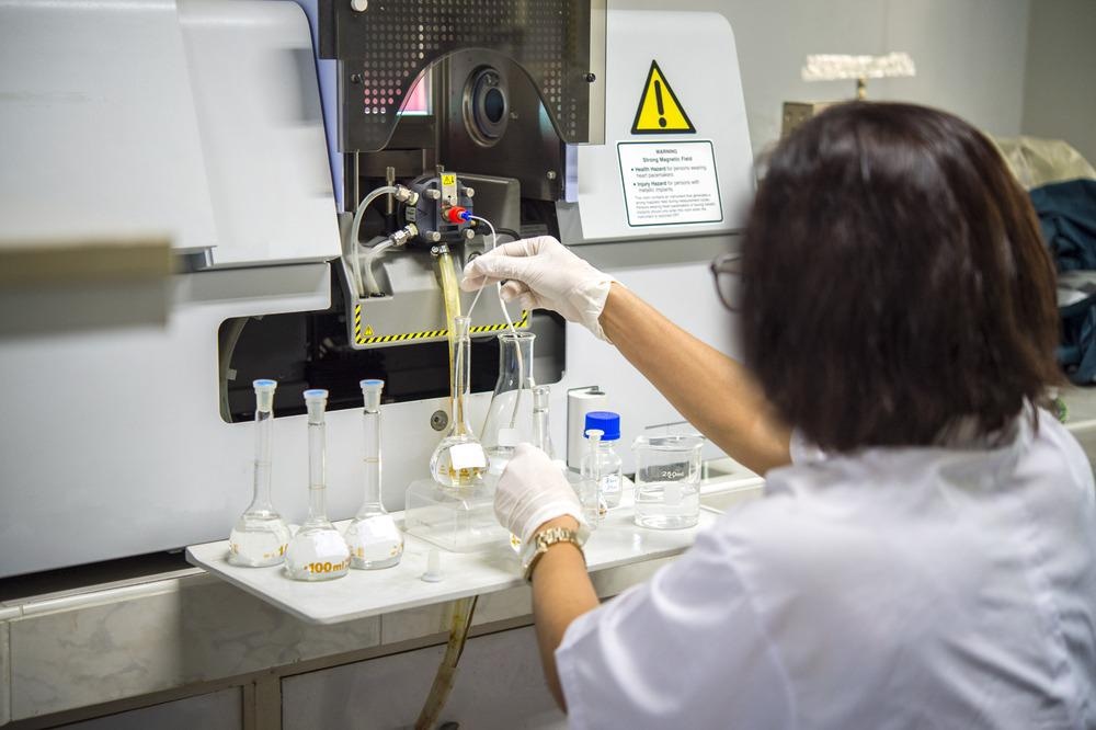
Raman spectroscopy is a powerful tool for analyzing a wide range of different materials. Since it was discovered, the technique has been employed to investigate material features such as carbonaceous and inorganic properties and their phases, functions, and defects.

Image Credit: Rabbitmindphoto/Shutterstock.com
Advances in instrumentation and techniques – particularly the development of surface and tip-enhanced Raman spectroscopy – have introduced Raman spectroscopy to the biological and analytical fields. Now, the robustness and versatility of the latest Raman instruments present a promising solution for on-field analysis of a range of materials.
What is Raman Spectroscopy?
Raman spectroscopy has opened the way for detailed knowledge of materials, particularly carbonaceous materials such as graphite, since it was established just a few decades ago. Now, Raman analysis has extended beyond the research laboratory, with industrial applications in sectors such as food and drug manufacturing, environmental monitoring, and textiles.
The technique was developed by two different scientists simultaneously in the early 20th century, without any knowledge of the others’ discoveries. Chandrasekhara Venkata Raman received a Nobel prize for his efforts, and the phenomenon and technique based on his observations were both named for him. But Grigorij Samuilovič came to the same conclusions as Raman independently, and at roughly the same time. Raman spectroscopy was widely established as an analysis method in the second half of the 20th century, once laser light-equipped spectrometers were first developed for research purposes.
Raman spectroscopy, like all spectroscopic methods, is based on the interactions between electromagnetic radiation and matter. Absorption, transmittance, and scattering are the phenomena governing interactions between energy and matter.
Scattering occurs when a photon meets a crystal lattice or molecule, distorting the cloud of electrons that surrounds its nucleus and changing its species polarization. Photons leave the system in a scattered pattern, which is either elastic (Rayleigh) or inelastic. In Rayleigh scattering, photons going into the material have a different energy to photons coming out.
The difference between incoming and outgoing photons’ energy is called Raman shift. Raman spectroscopy applies laser light to samples to generate this effect, then uses specialist sensors to record the resulting Raman spectrograph. Human or computer analyzers measure the Raman data from the spectrograph to characterize various target features in the sample.
In conventional Raman spectroscopy, spatial resolution is limited to around 1 mm2. Nanostructured materials are characterized using micro-Raman spectroscopy. This technique combines a Raman spectrometer with an optical microscope to focus the laser beam to approximately 1 μm. This enables scanning sample depths ranging from 1 μm down to just a few hundred nm.
Raman spectroscopy is one of the most versatile and robust non-destructive spectroscopy methods available to researchers and technologists. It is suitable both for lab- and field-based operations, and displays numerous advantageous features.
The presence of water, for example, barely affects the quality of signal collected. This means that Raman spectroscopy is suitable for many applications where infrared (IR) spectroscopy is not reliable.
Drawbacks include data analysis difficulties due to the massive datasets generated by Raman spectrometers. Highly active Raman species such as carbon particles may also mask the presence of other species in the sample. Research is underway to overcome these challenges.
Applications for Raman Spectroscopy
Raman spectroscopy has developed into a highly versatile and robust technique, with applications in materials science, biology, pharmacy, industry, and even on other planets.
The most well-established application for Raman spectroscopy is in carbon-based materials characterization, with the technique responsible for many discoveries in materials science in the last century.
Now, advanced carbonaceous materials such as carbon nanotubes (CNTs), graphene, and amorphous carbon are precisely measured and studied with Raman spectroscopy.
Raman spectroscopy is also employed in the study of metal, ceramic, and polymer nanoparticles. These materials may exhibit interesting properties for biological applications, including biocompatibility, magnetism, and photoluminescence. Characterizing them with Raman spectroscopy reveals these features, probing how they interact with the biological environment.
Biomolecules can also be directly sensed with Raman spectroscopy. This is enabling a new understanding of DNA, proteins, lipids, and carbohydrates in biological organisms.
In medicine, Raman spectroscopy is used to improve the accuracy of tumor diagnoses. Enchondroma and chondrosarcomas, thyroid cancer, colorectal cancer, prostate cancer, and many others have all been determined with Raman analyses of easily obtainable biological samples (serum, blood, saliva, urine, sperm, etc).
Raman analysis can also identify multiple pathologies and infections at once, which is especially important during pandemic events. As such, Raman spectroscopy has played an important role in the fight against COVID-19, identifying and characterizing the disease in a high quantity of samples at speed.
As it is non-destructive, precise, and quick, Raman spectroscopy is often used in mineralogy to analyze a large variety of samples effectively. It is often used to analyze jewels, minerals, works of art, and historical artifacts for these reasons. Cultural heritage diagnostics is a growing interdisciplinary field that unites the arts and the sciences, based around Raman spectroscopy results.
In industry, Raman spectroscopy is used for quality control, research and development, and environmental and waste monitoring.
Raman spectroscopy has even made its way into space. The Perseverance rover, which landed on Mars in February 2021, is fitted with two Raman instruments: SuperCam and SHERLOC (Scanning Habitable Environments with Raman and Luminescence for Organics and Chemicals).
References and Further Reading
Auer, B.M., and J.L. Skinner (2008) IR and Raman spectra of liquid water: Theory and interpretation. The Journal of Chemical Physics. https://doi.org/10.1063/1.2925258
Geraldes, C.F.G.C. (2020) Introduction to Infrared and Raman-Based Biomedical Molecular Imaging and Comparison with Other Modalities. Molecules. Available at: https://doi.org/10.3390/molecules25235547
Kudelski, A. (2008). Analytical applications of Raman spectroscopy. Talanta. Available at: https://doi.org/10.1016/j.talanta.2008.02.042.
Orlando, A., et al. (2021). A Comprehensive Review on Raman Spectroscopy Applications. Chemosensors. Available at: https://doi.org/10.3390/chemosensors9090262.
Raman, C.V. (1953). A New Radiation. Proceedings of the Indian Academy of Sciences. Available at: https://doi.org/10.1007/BF03052651.
Disclaimer: The views expressed here are those of the author expressed in their private capacity and do not necessarily represent the views of AZoM.com Limited T/A AZoNetwork the owner and operator of this website. This disclaimer forms part of the Terms and conditions of use of this website.