Neuroscientists can now watch neuron activity deep inside a living human brain thanks to a pioneering new optical technique. A team from the European Molecular Biology Laboratory (EMBL), based in Heidelberg, Germany, described the two-phase microscopy method for in vivo brain imaging in Nature Methods in September 2021.

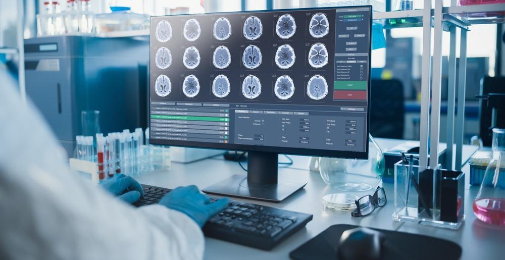
Image Credit: Gorodenkoff/Shutterstock.com
Breakthrough Combination Technique
The new technique combines discrete recent advances in microscopy and optics. 3-photon microscopy (3PEF) provides penetrative illumination and magnification. Advanced adaptive optics ensures a consistently high image quality by algorithmically optimizing the image data to minimize the effects of distortions.
The combination of these technologies creates a technique that can be used by neuroscientists to see deeper into a live brain – without harming the subject – and watch neuron activity as it happens. The technique brings a new level of high resolution to in vivo brain imaging.
The team behind this new development are members of Robert Prevedel’s leading research group focused on “advanced optical techniques for deep tissue microscopy.”
Challenges of In Vivo Brain Imaging
Visualizing neural cells in the deep region of the brain that looks after spatial memory and navigation, the hippocampus, has proved challenging for in vivo brain imaging.
Hippocampus phenomena such as “calcium waves,” astrocytes in deep layers of the cortex tissue, regularly occur in all live mammals’ brains. But, because they occur deep inside the brain’s structure, medical imaging techniques have been unable to provide significant usable data on how they work.
Traditional fluorescence brain microscopy used on ex vivo (dead) brain tissue samples does not provide the data needed by neuroscientists today, according to the EMBL researchers. It is also inadequate for traveling deep into tissue – where answers to many of the brain’s biological mysteries might lie.
Benefits of 3PEF
In traditional fluorescence microscopy, the fluorescence molecule absorbs two photons with each pulse. However, the further they have to travel, the more likely they are to scatter. Increasing the exciting photons’ wavelength towards the infrared helps to overcome this challenge by making sure radiation energy is absorbed in the fluorophore.
But 3PEF is even more effective at increasing penetration depth for medical imaging. The three photons used are less vulnerable to scattering and lead to higher definition images from deeper inside the tissue.
Utilizing Adaptive Optics
While 3PEF can provide a greater amount of image data, that data may not always be usable. Adaptive optics is applied as the second prong of the EMBL team’s two-phase technique to ensure images are not distorted, blurry, or otherwise damaged.
Adaptive optics is a technique borrowed from astronomy, in which algorithmically generated instructions tell computers how to replace pixels in image data to overcome known distortions. Atmospheric gases and gravity, for example, distort telescope images and have to be corrected.
The same is true on the quantum scale that 3PEF is designed to operate in. It is inhomogeneous tissue scattering rather than atmospheric gases that causes distortion in 3PEF, but adaptive optics can still be employed to fix this issue.
An actively controlled deformable mirror optimizes wavefronts to focus light deep inside the brain. The speed of this adaptive optics component had to be specially upgraded to enable it to be used for live-cell monitoring.
Benefits of the Combined Approach
Using 3PEF and adaptive optics together like this for the first time, the team showed the deepest ever in vivo images of live neurons at high resolution in its paper in Nature Methods. Dendrites and axons connecting neurons in the hippocampus were also visualized with the new technique.
The new technique also allowed for fewer measurements to be made to achieve high-quality images. This was a focus of the research, as it sought to minimize the invasiveness of in vivo brain imaging.
References and Further Reading
Booth, M.J. (2007). Adaptive optics in microscopy. Philosophical Transactions of the Royal Society A: Mathematical, Physical and Engineering Sciences. https://doi.org/10.1098/rsta.2007.0013.
EMBL’s Two-Phase Microscopy Technique Enables Deep In Vivo Brain Imaging. [Online] Optics.org. Available at: https://optics.org/news/12/10/3.
Kerr, J., and W. Denk (2008) Imaging in vivo: watching the brain in action. Nature Reviews Neuroscience. https://doi.org/10.1038/nrn2338.
Streich, L. et al. (2021) High-resolution structural and functional deep brain imaging using adaptive optics three-photon microscopy. Nature Methods. https://doi.org/10.1038/s41592-021-01257-6.
Disclaimer: The views expressed here are those of the author expressed in their private capacity and do not necessarily represent the views of AZoM.com Limited T/A AZoNetwork the owner and operator of this website. This disclaimer forms part of the Terms and conditions of use of this website.